Quantcast
Channel: Biosciences Biotechnology Research Asia
Viewing all 1376 articles
Browse latest View live

Association of ABO Blood Groups with Procrastination among Tuberculosis Patients in the Tribal Populations of Nagaland

$
0
0

Introduction

Tuberculosis (TB) remains a main public health problem in India, accounting for a quarter of the eight million instances of TB that arise worldwide. Despite the fact that tablets are available for remedy, TB stays nevertheless as a massive burden, in aid negative settings and the arena’s most vital motive of death specifically in India. When clinical knowledge is used to manual coverage and practices, evidences are ranked in line with the relative merits of various data.1 Over the decade, numerous new interventions in TB manipulate had been advanced and recommended in WHO hints and carried out into India’s TB manipulate programmed. In the northeastern states of India, TB and PRG are found to be endemic. While guidelines for brand spanking new interventions are normally based on proof from standard population, little is thought from tribal regions. According to Indian state TB statistics 2017 and 2018 it is found that Nagaland a North Eastern state of India inhibited by tribal population accounts to 5826 TB Patients Notified Public Sector out of 2,050,220 population. The Revised National Tuberculosis Control Programme (RNTCP) has adopted multiple strategies so as to reach overall coverage of tuberculosis diagnosis and treatment. However, rural populations such as those living in the hilly terrain of Nagaland have limited access to health structures due to “poor road conditions and lack of basic communication services”, which may also hinder completion of TB treatment20. Due to limited evidence, at the side of other factors, this could have hindered wide-scale use of tribal unique proof based interventions. One of the approaches is proof from it as a minimum one desirable systematic assessment. Its purpose is to reduce large portions of statistics to usable dimensions. Some declare that doing so is an effective medical approach; one of this is much less time ingesting and greater dependable then engaging in new research. The use and usefulness of systematic critiques is one effective mechanism for improving the proof available to tell population fitness decision making.1

Moreover they contend that the various occasions where in man or woman research is executed allow reviews consequences to be generalized throughout distinct contexts and emerge more extra tremendous than person research. There are currently rare systematically accumulated statistics on the availability of proof for scale-up of newly encouraged interventions for TB control in tribal areas.1

The polymorphism in the ABO blood group remains important in population genetic studies, estimating the availability of compatible blood, evaluating the probability of hemolytic disease in the new born, resolving disputes in paternity/ maternity and for forensic purposes2. The frequencies of ABO and Rh phenotypes in different populations have been extensively studied. Rh system emerged as second most important blood group system due to hemolytic disease of newborn and its importance in RhD negative individuals in subsequent transfusions once they develop Rh antibodies. The D antigen, after A and B, is the most important red cell antigen in transfusion practice3.

Several physical, emotional and mental problems appear to be associated with procrastination. It may create embarrassment and inferiority complex among students of which the found negative relationship between level of ego identity and procrastination; it lessens confidence among students and their expectancy of completing a task4; resulting in unhealthy sleep, diet and exercise habits 5 yields to higher rates of smoking, drinking, digestive ailments, insomnia and cold and& flu symptoms6; increases a lot of stress, worry, and fear leading a miserable life with shame and self-doubt creating and raising anxiety and deteriorates self-esteem7 affects achievement of goals creating anxiety8 and causes higher stress, low self-esteem, depression, cheating, plagiarism, higher use of alcohol, cigarette and caffeine and decreased ability to maintain healthy self-care habits like exercise and eating 9

The study was conducted with the objectives to the context of disease epidemiology and procrastination, upon various Indian tribal communities, who are highly isolated both demographically and topographically, from the mainstream Indian populations. Blood bank usually has a problem of ever-changing stock position and it being very difficult to predict the prevalence of a particular blood group at a particular time. The present study was done to assess the prevalence of blood groups in different categories of Northeast India with the following specific objectives;       (i) To evaluate the most common Blood group among tuberculosis patients (ii) To compare and study the controlled groups with those of tuberculosis patients.

Research Methodology

The present study was descriptive in nature. Therefore survey approach was considered and adopted to collect the data from respective respondents. 

Population and Sampling

The present study was delimited to the TB patients of the olive Christian Hospital, Dimapur, Nagaland. The population of the study is tuberculosis patients of the above said hospital with the ABO blood groups. Among the population 50 TB patients were selected as a sample through the random sampling technique Further, 50 control group respondents were selected from the faculty members of St. Joseph University to evaluate and compare the result. Overall 100 respondents were considered for the study including patients and control group.

Data Collection

The study was descriptive in nature; therefore, the researchers considering the survey approach was appropriate to adopt for data collection. The researchers has developed two different set of questionnaire for patient group and control group respectively based on 5 point Likert scale technique. And the developed an questionnaire has been validated through the pilot study with the 50 respondents. Some corrections were identified and rectified in the pilot study. Finally, the questionnaires were administered on TB Patients of olive Christian Hospital, Dimapur, Nagaland and Control group peoples. The collected data were coded and analyzed in terms of percentage and mean score through Ms-Excel.

Laboratory Analysis

Once the samples were collected the ABO and Rh blood group testing was done from the laboratory using the standard protocol of AB D Antisera typing Kit.

Statistical Analysis

The gene and allele frequencies of blood group, are calculated by Hardy-Weinberg model using S2 ABO estimator software.10 Allele Frequencies are calculated under the assumption of Hardy–Weinberg equilibrium and expressed as percentages. The chi – square test is used to compare observed allelic and genotypic frequency distributions of the blood group and Rh antigens to that of under the Hardy–Weinberg.11

Results and Discussion

Association studies between ABO blood groups with Procrastination among tuberculosis patient’s diseases are the hallmarks for unravelling the genetic pattern of complex diseases. Studying the relationship between allelic and genotype frequencies of candidate genes among both affected and healthy subjects, to understand the genetic etiology of complex human traits, is an efficient method to elucidate their disease pathogenesis. It is found that female occurred in the order O> A >B. The allele frequency of blood group O is the highest, 𝜒2 The goodness of fit test was resulted in value was = 5.1 and p value was 0.02. This only significant value(Table.2) There is the high proportion of Rh (D) +ve individuals than the Rh – ve in the study populations. (Table.1)

Table 1: Frequency of ABO blood groups and Rh factor in Patients and Controls

Group  A B AB O N Phenotypic

frequency

Rh+ Ve Rh-Ve
Control 9(18%) 11(22%) 0 30(60%) 50 O >B >A >AB 50
Male 4(14.8%) 10(37%) 0 16(59%) 27 O >B >A >AB 27
Female 5(21.7%) 2(7.4%) 0 16(69.5%) 23 O >A >B >AB 23
Case


16 (32%) 10 (20%) 0 24(48%) 50 O >A >B >AB 50
Male 4(16%) 3(12%) 0 18 (72%) 25 O >A >B >AB 25
Female 3   (12%) 1(4%) 0 21 (84%) 25 O >A >B >AB 25

Table 2: Shows the overall allele frequencies for the ABO in Patients and Controls

Group Group Gene frequency Hardy-

Weinberg log

likelihood


Genotypic

frequency

𝜒2 𝑃value
p[A] q[B] r[O]
Control Control 0.095 0.12 0.79 -48.6 O >B >A 1.4 0.23
Male 0.084 0.2 0.72- -33.5 O >B >A 0.001 0.97
Female 0.13 0.064 0.8 -22.88 O >A >B 1.05 0.3
Case


Case 0.185 0.12 0.7 -57.5 O >A >B 1.03 0.3
Male 0.1 0.07 0.82 -24.22 O >A >B 0.76 0.38
Female 0.08 0.02 0.88 -18.79 O >A >B 5.1 0.02*

* Statistically significant.

Hence forth, we tried to elucidate the possible association of the ABO blood groups with Procrastination among tuberculosis disease patients. The ABO blood groups with Procrastination were observed to be more prevalent among cases than controls, as shown in Table. 2 and 3. Genotype distributions, allelic frequencies and the corresponding odds ratios (OR) were calculated for each variant as shown in Table: 4.

Table 3: Distribution of ABO blood frequencies in Cases and Controls stratified according to gender

Sl.No Gender ABO blood Case

(n= 50)

Control

(n= 50)

χ2 OR 95% CI P-value
1. All (50) A 16 9 1.92 2.14 0.77-6.2 0.082
B 10 11 0.05 0.88 0.33- 2.32 0.5
O 24 30 1.0 0.615 0.27- 1.35 0.31
2. Female (20) A 2 5 0.69 0.33 0.056 – 1.97 0.45
B 0 2 0.5 0.0 0.00 – 3.43 0.46
O 18 13 2.29 4.8 0.86 –27.2 0.12
3. Male (30) A 4 4 0.41 1.0 0.22 – 4.43 0.70
B 8 10 0.08 0.72 0.23 – 2.20 0.77
O 18 16 0.07 1.32 0.77 – 3.65 0.79

χ2 : Chi-square with 1 degree of freedom; OR: odds ratio

No significant association was observed in the ungrouped data. This occurred in the order ABM (Dominant model). The allele frequency of blood group A +B Vs O is the highest, 𝜒2 The goodness of fit test was resulted in value was = 4.6 and p value was 0.05 this is significant value (Table. 4) However, when segregated the subjects into male and female, we found that the O Blood group homozygous genotype has a significant prevalence upon females than in males.

Table 4: Distribution of ABO blood frequencies (dominant and recessive model) in Patients and Controls

ABO blood Gender Model TEST Case

(n= 50)

Control

(n= 50)

OR 95% CI χ2 P-value
All A +B Vs O ABM 26 20 1.62 0.73 -3.57 1.0 0.31
B+O vs A REC1 34 41 0.4 0.17 – 1.19 1.92 0.16
A+O vs B REC2 40 39 1.12 0.42 – 3.02 0.0 0.8
Female A +B Vs O ABM 2 7 0.2 0.02 – 1.14 4.6 0.05*
B+O Vs A REC1 18 15 3.0 0.5 -17.7 1.51 0.21
A+O Vs B REC2 20 18 1.0 0.29 – -1.0 2.50 0.15
Male A +B Vs O ABM 12 14 0.76 0.27 -2.21 0.26 0.6
B+O Vs A REC1 26 26 1.0 0.22 – 4.43 0.0 1.0
A+O Vs B REC2 12 14 0.7 0.27 – 2.12 0.26 0.6

After the discovery of blood groups, numerous studies on associations of blood groups and various diseases were performed. Identifying the prognostic and associating factors, which predict the condition of the disease and its response to the treatment, can play an important role in determining the therapeutic strategies.

This study demonstrated that blood group O+ve was commonest and O-ve was least frequent among blood donors. This is in agreement with the studies that performed on blood donors12 and population of Tehran Province.13

Previous studies of the ABO pattern among patients with pulmonary tuberculosis were made at a time when the incidence was much higher than at present in Copenhagen. If a weak correlation exists between tuberculosis and some of the blood groups, it could easily have been obscured when the disease was more frequent. This is likely especially if recurrences, sequelae or non-bacillary patients were included. The ABO pattern of these patients is closer to or identical with the ABO pattern of normal persons. In this context it should be recalled that the diagnosis of pulmonary tuberculosis is less exact among abacillary patients. This fact may well explain the discrepancies in the ABO pattern of bacillary and abacillary patients, since the latter group may contain a number of patients not suffering from tuberculosis.

The present study showed that there was an association between tuberculosis and the blood groups B and AB in this region of the Dimapur City. The deviations were however not significant14.Many studies with similar intent were conducted earlier.

It is Suggested blood Groups O and AB individuals are more susceptible to TB15. However, a study by Rao et al., concluded that blood Groups O and A were the most common blood groups associated with PTB.16 A study in Gujarat, a significant association was discovered between blood Group AB and pulmonary TB.17 Similarly, Jain17 had similar observations for AB blood group and pulmonary TB. People with blood Group O showed protection from TB in a Chinese population.18This could be one of the most important for the observed deviations from the expected ABO pattern, but it remains speculative so far. Studies suggests that self-administered TB treatment is feasible for patients living in areas with limited or no access to health services.20

Conclusion

The study concluded that procrastination effects on the O +ve blood groups patients genotype is significantly associated more with the female patients (p = 0.02) than male patients (p = 3.8). The allele frequency of blood group O is the highest, 𝜒2 The goodness of fit test resulted in value was  5.1 and p value was 0.02  among tuberculosis patients in the tribal populations of  Nagaland. The significant association of the O blood genotype with Procrastination among tuberculosis patients, especially, with females of the tribal populations was observed.

The ABO blood groups with Procrastination were observed to be more prevalent among tuberculosis patients than the controls. When separated the subjects into male and female, it is found that the O Blood group homozygous genotype has a significant prevalence upon females than in males. With the observed association of ABO genotype, Procrastination with tuberculosis, this study anticipates more studies with larger cohorts to extend and elucidate. The in progress study be responsible for spirited information on the Procrastination of TB among the tribal population of  Nagaland which jerry can be situated used as a standard data for future epidemiological studies.

References

  1. V.G. RaoM. MuniyandiJ. BhatR. YadavR, “Sharma Research on tuberculosis in tribal areas in India,” A systematic Review , 2017, http://dx.doi.org/10.1016/j.ijtb.2017.06.001
  2. Bashwari LA, Al-Mulhim AA, Ahmad MS, Ahmed MA. Frequency of ABO blood groups in the Eastern region of Saudi Arabia. Saudi Med J, 2001; 22: 1008- 1012.
  3. Das PK, Nair SC, Harris VK, Rose D, Mammen JJ, et al. Distribution of ABO and Rh-D blood groups among blood donors in a tertiary care centre in South India. Trop Doct, 2001; 31: 47-48.
  4. Steel, P. Procrastination History, 2008. Retrieved from www.procrastinus-history.htm accessed on January 10, 2010.
  5. Sirois, F. & Pychyl, T. Academic Procrastination: Costs To Health And Well Being. Presentation at American Psychological Association Annual Convention, Chicago, August 22-25, 2002. Retrieved from www.prgtextbasedconferencesjumppage.html accessed on October 19, 2008.
  6. Akinsola , M. K., Tella, A. & Tella,A. Correlates of Academic Procrastination and Mathematics Achievement of University Undergraduate Students; in Eurasia Journal of Mathematics, Science & Technology Education, 2007, 3(4), 363-370.
  7. Hoover, E. The Chronicle Of Higher Education. Ohio State University, 2005, Retrieved fromhttp://www.physics.ohio-state.edu/-wilkins/writing/resources/essays/procrastination.html on September 15, 2008.
  8. Scher, S. & Nelson, L. Academic Procrastination: Affect, Achievement, Goals and Anxiety. Presentation at American Psychological Association Annual Convention, Chicago, August 22-25, 2002. Retrieved fromwww.prgtextbasedconferencesjumppage.html accessed on December 21, 2009.
  9. Goode, C. Effects Of Academic Procrastination: Students Procrastination Affects More Than Grades. 2008, Retrieved from website http://homeworktree.com/media/news-releases/academic-procrastination accessed on October 13, 2008.
  10. Pedro J.N. Silva Allele frequency estimation in the human ABO blood group System, 2002, http://alfl.cii.fc.ul.pt/¬pedro/Soft/ABOestimator/.
  11. Epi-Info-Community-Edition/license.md Github. Retrieved 18 January 2019.
  12. 12.Pourfathollah AA, Oody A, Honarkaran N. Geographical distribution of ABO and Rh (D) blood groups among Iranian blood donors in the year 1361(1982) as compared with that of the year 1380 (2001). Blood Journal, 2003 1(1): 11–19
  13. Farhud DD, Eftekhari A. Blood groups distribution in Iran. Iranian J Publ Health, 1994, 23(1): 1–10
  14. Rao BN, Reddy VD, Sahu PS, Veerendra Kumar A, David MA, Yugandhar P, et al. The ABO blood group distribution and pulmonary tuberculosis. J Clin Diagn Res 2012;6:943-6.
  15. Viskum K. The ABO and Rh blood groups in patients with pulmonary tuberculosis. Tubercle 1975;54:329-33.
  16. Gondaliya ST, Makwana HH, Lakum NR, Agnihotri AS. Pulmonary tuberculosis and blood groups: Any association? Gujarat Med J, 2012;67:39-41.
  17. Jain RC. ABO blood groups in relation to breast cancer. Curr Med Pract 1968;12:498.
  18. Saha N, Banerjee B. Incidence of ABO and RH blood groups in pulmonary tuberculosis in different ethnic groups. J Med Genet 1968;5:306-7.
  19. Mrinalini Das, Katerina Doleckova, Rahul Shenoy, Jagadish Mahanta, et.al. Paragonimiasis in tuberculosis patients in Nagaland, India. Glob Health Action. 2016; 9: 10.3402/gha.v9.32387.
  20. Mrinalini Das, Petros Isaakidis, Rahul Shenoy, Rey Anicete, Hemant Kumar Sharma, Imyangluba Ao et.al. Self-Administered Tuberculosis Treatment Outcomes in a Tribal Population on the Indo-Myanmar Border, Nagaland, India. PLoS One. 2014; 9(9): e108186.

Response for Callus Induction in Popular Indica Rice Varieties and Its Mutant Lines Using Different Media Combinations

$
0
0

Introduction

Rice is one of the prime food crops across the globe. It serves as the source of carbohydrate for more than half of the world’s population and it is cultivated under variety of agro-climatic conditions1.

Rice improvement through biotechnological approaches depend on an efficient protocols through, in vitro callus induction as well as a suitable regeneration protocols2-4. In vitro rice regeneration can be accomplished through somatic embryogenesis and organogenesis. Somatic embryogenesis of rice is one of the most promising approaches for rapid propagation due to production of large numbers of plantlets and for the application of genetic transformation technology especially against biotic stresses5. The success of somatic embryogenesis is highly influenced by a suitable genotype, growth medium, plant growth regulators (PGRs), carbon sources and gelling agent affect the somatic embryogenesis as well as plant regeneration of rice6.

But, most of the indica rice genotypes are recalcitrant for tissue culture studies, i.e., they exhibit poor response for callus induction and plant regeneration7. Callus induction potential of a particular genotype is fundamental requirement for tissue culture based crop improvement, especially for genetic transformation8. Hence, a study was conducted in order to optimise the callus induction protocol of popular indica genotypes for its utilization in tissue culture based crop improvement programmes.

Materials and Methods

In Vitro Response of Different Genotypes to Callus Induction Ability

The response of popular rice genotypes for their difference in callus induction ability was studied under controlled in vitro condition. An elite indica cultivar, BPT 2231 (Akshaya) was used for optimisation of callus induction media. Then the appropriate five different concentrations of media were used for other indica rice genotypes that were used in this study. The protocol for callus induction and various genotypes used in the study were described in this section and in Table 1.

Well-filled, mature and dehulled seeds were taken for callus induction studies. They were washed thoroughly with running tap water for ten minutes to wash off external dust or contaminants. Later the explant materials were washed with Tween®20 detergent solution (Sigma-Aldrich chemicals Pvt. Ltd.) for 15 minutes. To remove the detergent the explant materials are thoroughly washed several times with sterile water. The explants are then surface sterilized in Laminar Airflow Chamber (Cleanair Flow Systems, India).

Surface Sterilization of Explants

After preliminary washings, the explants were taken to laminar air flow chamber and surface sterilization was done by keeping the explants for three minutes in 70% ethyl alcohol (Changshu Hangsheng Fine Chemicals Co., Ltd. China) followed by treatment with 0.1% mercuric chloride (HiMedia Laboratories, LLC) for eight to ten minutes9 and finally washed thoroughly with sterile distilled water for 3-5 times to remove any traces of mercuric chloride.

Two to three seeds per tube were inoculated in the media for callus induction and kept in darkness at 25ºC with a lightly intensity of 2000-3000 lux alternatively maintained at a cycle of 16 hours light and 8 hours dark for three weeks.

Table 1: List of genotypes used for Tissue culture Studies

S. No. Genotypes Special Features Source
1 BPT 2231 Low glycemic index Rice Research Unit, Bapatla
2 CR 1009 Resistant to BPH Dept. of Farm management, AC&RI, Madurai
3 CR 1009 Sub1 Submergence tolerant Dept. of Farm management, AC&RI, Madurai
4 CO 52 Medium slender grains Dept. of Farm management, AC&RI, Madurai
5 ADT 45 Resistant to Gall midge Dept. of Plant Breeding and Genetics, AC&RI, Madurai
6 ADT 37 Multiple resistance to many pests and diseases
7 M6 Electron beam mutant of ADT 45
8 M55 Electron beam mutant of ADT 45
9 M66 Electron beam mutant of ADT 45
10 M78 Electron beam mutant of ADT 37
11 M79 Electron beam mutant of ADT 37
12 M85 Gamma ray mutant of ADT 37
13 M88 Electron beam mutant of ADT 37

Different Media Composition for Callus Induction Studies

Murashige and Skoog (MS) medium10 was chosen for the present investigation, along with different hormonal combinations of 2,4-Dichlorophenoxy acetic acid (2,4-D) and kinetin (KN). The different media combinations are described in Table 2.

Table 2: Different media combinations used in the study

Treatment Basal Media PGR (mgl-l)
2,4-D KN
C1 MS 2 1.5
C2 MS 1.5 2
C3 MS 2 2
C4 MS 2.5 2
C5 MS 2 2.5

 Five replications were maintained in each media combinations and 50 explants were maintained in each of the replication. Series of events during callus induction is presented in Figure 1. The callus induction ability of a genotype is a complex trait that denotes both quantity and quality of the callus. Five different traits viz., Number of days for callus induction, callus induction percent, fresh weight of callus, dry weight of callus, percent of white/yellow callus and percent of callus browning were hence observed in this study for the precise estimation of the callus induction ability of a genotype.

Figure 1: Series of events during callus induction Figure 1: Series of events during callus induction

Click here to View Figure

 Statistical Analysis

The data was analysed in Factorial Completely Randomised Design (FCRD) using AGRES statistical software and the treatmental means were grouped using Least Significant Difference (LSD) method.

Results and Discussion

The effect of genotypes and different media combinations on various traits observed on callus was presented in Table 3.

Table 3: Response of genotypes and media combinations on various traits observed on callus

Treatment DCI(Days) CIP (%) CFW(g) CDW(g) WYC (%) CBW (%)
BPT 2231 17.16c 70.56b 0.3530a 0.0170f 60.24c 10.32d
CR 1009 15.28b 81.44a 0.2886c 0.0220e 76.64ab 4.80a
CR 1009 Sub1 17.00c 81.92a 0.2613d 0.0222de 75.12b 6.80bc
CO 52 14.08a 83.68a 0.1949h 0.0288a 78.40a 5.28ab
ADT 37 20.28d 60.16d 0.2116g 0.0276b 50.80ef 9.36d
ADT 45 20.40d 61.52d 0.2440e 0.0279ab 51.76e 9.76d
M6 22.28g 45.60h 0.1299j 0.0142h 22.48i 23.12f
M55 21.92gf 57.28e 0.1398j 0.0156g 37.36g 19.92e
M66 20.28d 51.20g 0.1590i 0.0175f 31.36h 19.84e
M78 20.52de 54.32f 0.2288f 0.0222de 48.48f 5.84abc
M79 20.28d 66.00c 0.2844c 0.0247c 60.08c 5.92abc
M85 21.52efg 64.16c 0.3272b 0.0231d 56.88d 7.28c
M88 21.16def 59.60de 0.3357b 0.0179f 53.44e 6.16abc
C1 18.98c 65.26c 0.2299c 0.0222c 55.29c 9.97c
C2 13.63a 72.49b 0.2852b 0.0251b 63.75b 8.74b
C3 15.46b 77.78a 0.3666a 0.0296a 70.55a 7.23a
C4 25.97e 50.06e 0.1466e 0.0145e 36.95e 13.11d
C5 22.94d 56.49d 0.1865d 0.0166d 43.85d 12.65d

DCI- Days to callus induction; CIP- Callus induction percent; CFW- Callus fresh weight; CWD- Callus dry weight; WYC- Percent of White/ Yellow callus; CBW- Percent of callus browning

Values with same alphabets are not significantly different

Callus induction ability of different genotypes studied in different growth media exhibited significant variability for all the traits.

The variety CO 52 was found superior in callus induction ability among other genotypes. It out-performed other genotypes in most the traits studied viz., number of days for callus induction (14.08), callus induction percent (83.68), dry weight of callus (0.0288) and percent of white/yellow callus (78.40). The mutant M6 was found to be the poorest performing genotype and it produced poor quality callus in all the treatments. The traits, number of days for callus induction (22.28), callus induction percent (45.60), fresh weight of callus (0.0299), dry weight of callus (0.0142) and percent of white/yellow callus (22.48) and percent of callus browning (23.12), were all found to be inferior compared to other genotypes.

MS medium is considered as the most suitable culture medium for callus induction of rice cultivars11, 12. Previous reports have also shown high callus induction on MS medium supplemented with 2,4-D13. Also, some reports suggest that the efficiency of callus induction is increased in a media containing proportionate quantity of both auxin and cytokinin14.  Similarly, among the different media combinations, MS + 2 mgl-l 2,4-D + 2 mgl-l KN produced high quality callus in most of the traits studied, while MS + 2.5 mgl-l 2,4-D + 2 mgl-l KN was found to be poor media for callus induction of different genotypes. The difference in callus formation between different media combinations are presented in Figure 2.

Figure 2: Difference in callus induction potential in different media combinations. Figure 2: Difference in callus induction potential in different media combinations.

Click here to View Figure

Differences in Callus Induction Ability among Mutants

Varietal differences in callus induction potential of rice genotypes have already been reported by several authors15, 16. The treatmental means were grouped by LSD method using AGRES statistical software and the differences among the mutants were studied and presented herewith.

Number of days for callus induction for M66 was found to be on par with its parent ADT 45 and M79 found to be on par with its parent ADT 37. Other studied mutants exhibited significant differences in mean values compared to parents and fall into other groups also.

Callus induction percent of all the mutants were statistically different compared to their parents. However, some of the mutants (M79 and M85) were grouped together or they were said to be on par, producing similar callus induction percent. Previous reports also showed the effect of rice genotypes on the variability of callus induction potential17, 18. In a similar study involving 510 indica and japonica accessions19, they found significantly different callus induction potential between the categorized populations.

Fresh weight of callus also showed significant differences in their mean value among the mutants to their parents. However, some of the mutants were grouped together (M6 and M55; M85 and M88) indicating absence of statistical significance between them. Similarly, dry weight of callus also exhibited significant differences in mean among the mutants, compared to their parents.

Percent of White/Yellow callus in M78 was found to be on par with its parent, while all other mutants exhibited significant difference in mean to their parents. However, in case of percent of callus browning, all the mutants studied exhibited significant difference in mean to their parents. However, they exhibit some amount of statistical similarity among them (M55 and M66; M78, M79 and M88). This result clearly indicated the genetic variability of rice cultivars with regard to callus browning which corresponds with the earlier reports20.

Conclusion

The response of different genotypes for callus induction indicates their potential to use them in a genetic engineering programme for transformation. Further, the best callus induction medium, MS + 2 mgl-l 2,4-D + 2 mgl-l KN, for most of the traits under study, was identified. The different mutants were also included in the study to estimate the differences in callus induction potential between its wild type. The significant differences found in callus induction potential may be due to somaclonal variation occurred in the particular gene. Hence, further studies in these mutants and its wild type can be used for identification of candidate genes controlling the traits studied.

Acknowledgement

The authors extend our gratitude to Department of Plant Breeding and Genetics, Agricultural College and Research Institute, Madurai for providing facilities to carry out the research work.

 Conflict of Interest

The authors have declared that no competing or conflicts of interest exist.

Funding Source

No receipt any financial support for the research

References

  1. Tyagi A.K., Khurana J.P., Khurana P., Raghuvanshi S., Gaur A., Kapur A., Gupta V., Kumar D., Ravi V., Vij S. Structural and functional analysis of rice genome. J. Genet. 2004;83(1):79-99.
  2. Khatun R., Islam S., Ara I., Tuteja N., Bari M. Effect of cold pretreatment and different media in improving anther culture response in rice (Oryza sativa L.) in Bangladesh. Indian J. Biotechnol. 2012;11:458-463.
  3. Siddique A.B., Ara I., Islam S.S., Tuteja N. Effect of air desiccation and salt stress factors on in vitro regeneration of rice (Oryza sativa L.). Plant Signal. Behav. 2014;9(12):e977209-1-e977209-10.
  4. Vega R., Vásquez N., Espinoza A.M., Gatica A.M., Valdez-Melara M. Histology of somatic embryogenesis in rice (Oryza sativa cv. 5272). Rev. Biol. Trop. 2009;57:141-150.
  5. Zuraida A., Naziah B., Zamri Z., Sreeramanan S. Efficient plant regeneration of Malaysian indica rice MR 219 and 232 via somatic embryogenesis system. Acta Physiol. Plant. 2011;33(5):1913-1921.
  6. Deo P.C., Harding R.M., Taylor M., Tyagi A.P., Becker D.K. Somatic embryogenesis, organogenesis and plant regeneration in taro (Colocasia esculenta var. esculenta). Plant Cell Tiss. Org. Cult. 2009;99(1):61-71.
  7. Ngezahayo F., Dong Y., Liu B. Somaclonal variation at the nucleotide sequence level in rice (Oryza sativa L.) as revealed by RAPD and ISSR markers, and by pairwise sequence analysis. J. Appl. Genet. 2007;48(4):329-336.
  8. Carsono N., Yoshida T. Identification of callus induction potential of 15 Indonesian rice genotypes. Plant Prod. Sci. 2006;9(1):65-70.
  9. Padhi M., Singh S. Surface sterilization for reducing microbial contamination in in vitro propagation of lasora (Cordia myxa Roxb.) using nodal segments. Int. J. Curr. Microbiol. Appl. Sci. 2017;6(8):836-842.
  10. Murashige T., Skoog F. A revised medium for rapid growth and bio assays with tobacco tissue cultures. Physiol. Plant. 1962;15(3):473-497.
  11. Azria D., Bhalla P.L. Plant regeneration from mature embryo-derived callus of Australian rice (Oryza sativa L.) varieties. Aust. J. Agric. Res. 2000;51(2):305-312.
  12. Niroula R., Sah B., Bimb H., Nayak S. Effect of genotype and culture media on callus induction and plant regeneration from matured rice grain culture. J. Inst. Agric. Anim. Sci. 2005;26:21-26.
  13. Chauhan M., Kothari S. Optimization of ionic and chelated iron and its interaction with disodium ethylenediaminetetraacetic acid for enhancement of plant regeneration in rice (Oryza sativa L). J. Plant Biochem. Biot. 2004;13(1):33-37.
  14. Trunjaruen A., Sayam R., Maneerattanarungroj P., Taratima W. Effects of Cultivation Media on In vitro Callus Induction and Regeneration Capabilities of Pakaumpuel Rice (Oryza sativa L.), Thai Rice Landrace. Walailak J. Sci. & Tech. 2020;17(1):37-46.
  15. Paul S., Roychoudhury A. Comparative Analyses of Regeneration Potentiality of Eight Indigenous Aromatic Indica rice (Oryza sativa L.) Varieties. Int. J. Sci. Res. Biol. Sci. 2019;6(1):55-64.
  16. Rima K., Pankaj K., Sharma V., Harsh K. Effect of culture media on seed germination and callus induction from cultured seeds of rice cultivars. Res. J. Biotechnol. 2020;15(3):33-40.
  17. Iqbal M., Hasan M., Ali M., Mian M., Hossain M., Ahmed m. In Vitro Callus Induction and Plant Regeneration Capacity Of Six Aromatic Rice Varieties. Intl. J. BioRes. 2013;15(4):55-60.
  18. Roly Z.Y., Islam M.M., Shaekh M.P.E., Arman M.S.I., Shahik S.M., Das D., Haamem M.M.E., Khalekuzzaman M. In vitro callus induction and regeneration potentiality of aromatic rice (Oryza sativa L.) cultivars in differential growth regulators. Int. J. Appl. Sci. Biotechnol. 2014;2(2):160-167.
  19. Zhang Z., Zhao H., Li W., Wu J., Zhou Z., Zhou F., Chen H., Lin Y. Genome‐wide association study of callus induction variation to explore the callus formation mechanism of rice. J. Integr. Plant Biol. 2019;61(11):1134-1150.
  20. Ogawa T., Fukuoka H., Yano H., Ohkawa Y. Relationships between nitrite reductase activity and genotype-dependent callus growth in rice cell cultures. Plant Cell Rep. 1999;18(7-8):576-581.


Biological Effect on Adhatoda Vasica Extract and its Combination with Antibiotics

$
0
0

Introduction

Nature gives several things against several diseases since last many decades. (Samuelsson et al., 2017) Adhatoda Vasica is a perpetual plant having a place with family Acanthaceae, generally known as Vasaka. (Claeson UP, et al., 2000) It is a small evergreen, perpetual bush dispersed all through India. It has been utilized for the treatment of different infections and clutters, especially for the respiratory tract illnesses. It is recognized with an upsetting smell and bitter taste. (Maurya S, et al., 2010) Vasicine and vasicinone alkaloids are the significant chemical constituents of the plant and are liable for its solid respiratory energizer activity. (Rastogi RP, et al., 1999) The drug for the most part contains fresh or dried leaves however the flowers, foods grown from the ground are likewise broadly utilized for different illnesses. Different measurement types of leaf-like powder, fresh juice, decoction, alcoholic and aqueous extract, and so on., are portrayed to be utilized for different diseases. (Anonymous ,1985)

A.Vasica was especially utilized as a home-grown medication for treating respiratory grievance including cold, hack, incessant bronchitis, asthma and as antispasmodic. (Singh B, et al., 2013) It has been accounted for antibacterial, injury mending, hypoglycemic, abortifacient, antitussive, anti-inflammatory and hepatoprotective activities. (Ahmad S, et al., 2009) Most part contains phytochemical likes pyrroquinazoline alkaloids, flavonoids, triterpenes, (Atta-Ur-Rahman, et al., 1997) steroids, tannins, saponins and glycosides. (Yusuf M, 2016) Therefore, influencing the bacterial pathogenic potential through majority detecting restraint is one of the new methodologies that are utilized to battle the microbial resistance. (Bhardwaj K, et al., 2013)     In laboratory experiments on cats and dogs, A. Vasica increases bile activity when animals are given a dose of 5mg / kg. in dogs, the amount of excreted bile increases by 40-100%. Animals also showed increased bilirubin excretion. (Gangwar et al. 2014)

In recent time medicine in surgery, cancer chemotherapy, and limb transplantation is credited to the use of antibiotics. (Livermore et al., 2002) Antibiotics working today will not work tomorrow New drugs must be examined, with fewer resistance (Sarkar et al., 2003) As a resistance to the spread of old antibiotics, new immune agents are rapidly developed. Yet, records of the rapid emergence and emergence of new antimicrobial agents suggest that a new family of antimicrobial agents will have a shorter life span. (Coates et al., 2002) For that much chemical contained the Adhatoda Vasica is effective against serval disease. Combination of plant extracts and antibiotics were used to increase sensitivity of plant extract and lower side effect of allopathy tablets.

Common Name
English : Malabar nut, Adulsa
Hindi : Adosa, Adalsa, Vasaka
Gujarati :  Ardusi, Adusi
Marathi : Vasuka

Plant Anatomy (Flora of Gujarat state, 1978)
Kingdom :   Plantae
Division  :  Angiosperm
Class        : Dicotyledonae
Series       :  Bicarpellatae 
Order       :  Personales
Family     :  Acanthaceae
Genus      :  Adhatoda
Species    : Vasica

Bio_Jiga_17_2_Fig1 Figures 1: Adhatoda Vasica Plant

Click here to View Figure

Materials and Methods

Material Use for Study              

Adhatoda Vasica Plant

Antibacterial Antibiotics :   Amoxicilline,   Ciprofloxacin, Ceftazidime, Erythromycin
Antifungal Antibiotics :   Amphotericin-B, Fluconazole
Bacteria  :   Bacillus subtilis (MTCC 441), Escherichia coli (MTCC 1687), Pseudomonas aeruginosa (MTCC 1688), Staphylococcus aureus (MTCC 737)
Fungi :  Aspergillus Niger (MTCC 1344), Candida albicans (MTCC 81)

Method

Leaves of the Adhatoda Vasica was conglomerate from local area of north Gujarat in June 2018. Identification of the plant was done by Dr. N. K. Patel, head of the botany department, Sheath M. N. Science college, Patan.  The first stages of the study of medicinal plants are to prepare plant samples, which is to store biomolecules in plants before extracted. The leaves were thoroughly wash with tap water and then washed with deionized water to remove particles of dust and sand. The leaves were dried in the dark at room temperature for several days and then powder with mortar. And it is extracted by using Soxhlet extraction.

Qualitative Phytochemical Analysis

Following methods were used for analysis of different type phytochemicals.

Test for Alkaloids

Plant Extractions were dissolved in dil. HCl and filtered

Dragendroff’s Test

Plant extract   +   Potassium Bismuth Iodide solution (Dragendroff’s Reagent)   →   Orange Precipitate

Hagers’s Test

Plant extract   +   Picric Acid (Hagers’s Reagent)       Yellow Precipitate

Mayers’s Test

Plant extract   +   Potassium Mercuric Iodide solution (Mayers’s Reagent)      Cream precipitate

Wagner’s Test

Reaction of plant extract   +   Iodine in Potassium Iodide (Wagner’s Reagent)      Red-brown precipitate

Test for Flavonoids

Test with Alkaline

Plant extract   +   Sodium Hydroxide      intense yellow color

which become colorless on further adding Dil. acid

Test with Lead Acetate

Treat plant extract   +   few drops of Lead Acetate      Yellow color precipitate

Shinoda Test

Dried powder of plant   +   5 ml of 95% CH3OH   +   Few drops of conc. HCl along   +   0.5 g magnesium      turnings Pink precipitate

Test for Glycosides

Keller-Kiiani Test

Plant extract   +   gla. CH3COOH    +   few drops 5% FeCl3   +   concentrated sulfuric acid       formation of   blue color in acetic layer.

Legal Test

Dissolve plant extract in pyridine   +   sodium nitroprusside solution   +   made alkaline       Pink or red Color produce

Test for Phenolics

FeCl3 Test

Plant extract   +   aqueous FeCl3       blue color

Test for Saponins

Foam Test

Plant extract in test tube with a little quantity of water       foam produced persisted for 10 minutes.

Test for Tannins

Gelatin Test

Plant extract   +   1% Gelatin Solution containing 10% sodium chloride       White precipitate

FeCl3 Test

Plant extract   +   FeCl3       Blue-black precipitate

Test for Terpenoids

Salkowski Test

Extract   +   Con. H2SO4       formation of yellow colored at lower layer

Preparation of Combination

Activities of various extract and their combination were resolute by the zone of ​​the inhibition method. It was investigated by agar diffusion method using agar cup method. Purified extract was diluted in dimethyl sulfoxide and purified antibiotics were used by purification; and stand at 40 C.  For compare slandered results we used standard antibiotic. Antimicrobial activity of all extracts and their combination were tested against for some bacteria and fungi which was prepared in Acetone (CH3COCH3), Methanol (CH3OH) and ethanol (CH3CH2OH) solvents. Muller-Hinton agar plates were seeded with indicator bacterial and fungal strains, and incubated for 24 hours at 370 C. The sensitivity of microbial species to plant extracts was determined by measuring the resistance zone size on the agar surface.

Result and Discussion

Table 1: Qualitative Phytochemical Analysis

Phytochemical Name of Test Acetone extract Ethanol Extract Methanol Extract 
Alkaloids Dragendroff’s Test + +
Hagers’s Test + +
Mayers’s Test + +
Wagner’s Test + +
Flavonoids Test with Alkaline + +
Test with Lead Acetate + +
Shinoda test + +
Glycosides Keller-Kiiani Test +
Legal Test + +
Phenolics FeCl3 Test + + +
Saponins Foam Test + +
Tannins Gelatin Test
FeCl3 Test + + +
Terpenoids Salkowski Test + + +

(+) shows the Positive result and (-) show Negative Result of the Test

Antimicrobial Analysis Adhatoda Vasica, Antibiotics and Combination both for 25 μl.

*Growth of the Bacteria and Fungi shown in table number 2 to table number 9 are in Millimeter (mm)

Table 2: Antibacterial activity of Adhatoda Vasica


Bacteria


Ac extract EtOH extract MeOH extract
Concentration (µg/ml)
1000 500 250 125 1000 500 250 125 1000 500 250 125
S. A 8 7 6 5 9 8 7 6 10 9 8 7
B. S 7 6 6 5 8 7 7 6 9 8 8 7
P. A 11 10 9 8 12 11 10 9 13 12 11 10
E. C 13 12 11 10 14 13 12 11 15 14 13 12

In the above table three different solvent were used for biological study. The study for each solvent were done with four different concentration. Table shows methanolic extract of in 1000 µg/ml concentration gives highest growth Which is used to decide dosage for human body.

Table 3: Antibacterial activity of Adhatoda Vasica with Amoxicilline

Bacteria


Amo Ac extract + Amo EtOH extract + Amo MeOH extract + Amo
Concentration (µg/ml)
1000 500 250 125 1000 500 250 125 1000 500 250 125
S. A 28 30 29 29 28 31 30 29 29 32 31 30 29
B. S 30 28 28 27 26 30 30 29 28 31 31 26 25
P. A 1 11 9 9 8 13 11 10 9 14 12 11 10
E. C 1 14 13 11 10 14 13 12 10 16 14 13 12

Amoxicilline which is an antibacterial antibiotic is combined with the plant. The study shows the combination with 1000 µg/ml in MeOH extract gives highest zone of inhibition.

Table 4: Antibacterial activity of Adhatoda Vasica with Ciprofloxacin

Bacteria


Cip Ac extract + Cip EtOH extract + Cip MeOH extract+ Cip
Concentration (µg/ml)
1000 500 250 125 1000 500 250 125 1000 500 250 125
S. A 24 26 25 25 24 30 29 27 22 29 28 27 26
B. S 27 29 28 27 26 30 29 27 27 31 30 29 28
P. A 27 33 31 30 28 35 32 30 29 36 35 32 30
E. C 27 32 30 29 28 34 32 32 28 35 33 31 30

 The mixture of Plant extract and ciprofloxacin in 1000 µg/ml gives good values in zone of inhibition of bacteria.

Table 5: Antibacterial activity of Adhatoda Vasica with Ceftazidime

Bacteria   Cef Ac extract + Cef EtOH extract + Cef MeOH extract + Cef
Concentration (µg/ml)
1000 500 250 125 1000 500 250 125 1000 500 250 125
S. A 1 9 7 6 5 10 8 7 5 12 10 9 7
B. S 1 8 6 6 5 9 7 7 6 10 8 7 6
P. A 5 13 11 10 9 16 14 12 10 19 17 14 13
E. C 16 22 20 19 18 26 25 23 21 27 24 22 19

Ceftazidime which is also an antibacterial antibiotic also gives effective results in methanol when it mixed with plant 

Table 6: Antibacterial activity of Adhatoda Vasica with Erythromycin

Bacteria  Ery Ac extract +Ery EtOH extract + +Ery MeOH extract +Ery
Concentration (µg/ml)
1000 500 250 125 1000 500 250 125 1000 500 250 125
S. A 16 22 22 21 19 22 21 24 23 23 21 20 19
B. S 22 24 24 23 21 26 25 24 23 25 24 23 21
P. A 1 13 11 9 8 14 11 10 9 15 13 11 10
E. C 5 17 17 16 15 19 17 17 16 19 17 16 15

Combination of plant extract and an antibacterial antibiotics Erythromycin gives good activity in higher concentration.

Table 7: Antifungal activity Adhatoda Vasica

Fungi  Ac extract EtOH extract MeOH extract
Concentration (µg/ml)
1000 500 250 125 1000 500 250 125 1000 500 250 125
A. N 8 7 6 5 9 8 7 6 10 9 8 7
C. A 7 6 5 4 9 7 5 4 9 8 7 5

In this table antifungal activity of Adhatoda Vasica extract was studied in Acetone, Methanol and Ethanol solvent. Biological study in two fungi, Aspergillus Niger and Candida Albicans fungi shows increasing of zone inhibition in higher concentration.  Methanolic extract shows higher   activity then other two solvent.

Table 8: Antifungal activity of Adhatoda Vasica with Amphotericin B

Fungi Amp Ac extract +Amp EtOH extract +Amp  MeOH extract +Amp
Concentration (µg/ml)
1000 500 250 125 1000 500 250 125 1000 500 250 125
A. N 14 19 19 18 18 19 17 16 14 22 21 20 19
C. A 8 12 10 9 8 18 16 13 12 19 17 14 13

An antifungal antibiotic Amphotericin B when combined with Adhatoda Vasica extract gives very good activity in 1000 µg/ml concentration.

Table 9: Antifungal activity of Adhatoda Vasica with Fluconazole

Fungi  Flu Ac extract + Flu EtOH extract + Flu MeOH extract + Flu
Concentration (µg/ml)
1000 500 250 125 1000 500 250 125 1000 500 250 125
25 µl 25 µl 25 µl 25 µl 25 µl 25 µl 25 µl 25 µl 25 µl 25 µl 25 µl 25 µl 25 µl
A. N 1 9 9 8 8 10 8 7 6 12 11 10 8
C. A 1 7 6 5 4 9 7 5 4 9 8 7 5

Fluconazole which is also an antifungal antibiotic also gives effective activity in higher concentration of the solvent.

Figure 2: Activity in Adhatoda Vasica extract (a) Antibacterial (b) antifungal Figure 2: Activity in Adhatoda Vasica extract (a) Antibacterial (b) antifungal

Click here to View Figure

Conclusion

Plant drugs from Ayurvedic framework are being investigated globally. The consequences of the essential examination demonstrated that alongside its traditional helpful cases and some experimentally demonstrated pharmacological exercises, Adhatoda Vasica additionally has the possibility to be created as a powerful plant with combination of antibiotics. Combination of A. Vasica and antibiotics indicated pronounced antimicrobial activities with wonderful restraints. This work gives logical confirmations to medicinal uses of A. Vasica with commitment of a portion of the distinguished and tried phytoconstituents in the got organic impacts. From the study we find that the combination of plant extract with antibiotics gives very effective activity then only plant and antibiotics. It also suggests that methanolic extract with higher concentration gives best results than others.

Acknowledgement

The authors are thankful to the Faculty member of Chemistry Department of Sheth M. N Science College, Patan. We are also thankful to the Microbiology Department of Dr. Indu Dayal Meshri College of Science and Technology, Patan for their kind support during the biological Study. We are also thankful to Dr. N. K. Patel, Head of Botany Department, Sheth M. N. Science College, Patan for identification of plant

Conflict of Interest Statement

We, the authors of the submitted manuscript declare that the work and data present in the manuscript entitled – qualitative analysis and biological study is genuine research carried out by us. The work finally belongs to the institutes. We have not misused the data previously published and have not manipulated the original work.

References

  1. Ahmad S, Garg M, Ali M, Singh M, Athar MT, Ansari SH. A phyto-pharmacological overview on Adhatoda zeylanica Medic. syn. A. vasica (Linn.) Nees, 2009.
  2. The Wealth of India, Council of Scientific and Industrial Research, New Delhi, India. 1985; 76.
  3. Atta-Ur-Rahman, Sultana N, Akhter F, Nighat F, Choudhary MI. Phytochemical studies on Adhatoda vasica Nees. Natural Product Letters. 1997 Oct 1;10(4):249-56.
  4. K Bhardwaj A, Vinothkumar K, Rajpara N. Bacterial quorum sensing inhibitors: attractive alternatives for control of infectious pathogens showing multiple drug resistance. Recent patents on anti-infective drug discovery. 2013 Apr 1;8(1):68-83.
  5. Claeson UP, Malmfors T, Wikman G, Bruhn JG. Adhatoda vasica: a critical review of ethnopharmacological and toxicological data. Journal of Ethnopharmacology. 2000 Sep 1;72(1-2):1-20.
  6. L Shah., Flora of Gujarat state. 1978, 1,48.
  7. Gangwar AK, Ghosh AK. Medicinal uses and pharmacological activity of Adhatoda vasica. International journal of herbal medicine. 2014;2(1):88-91.
  8. Livermore DM. Antibiotic resistance in staphylococci. International journal of antimicrobial agents. 2000 Nov 1;16:3-10.
  9. Maurya S, Singh D. Quantitative analysis of total phenolic content in Adhatoda vasica Nees extracts. International Journal of PharmTech Research. 2010 Oct;2(4):2403-6.
  10. Rastogi RP, Mehrotra BN. Compendium of Indian medicinal plants Vol. 2. Central Drug Research Institute; 1999,14.
  11. Samuelsson G, Bohlin L. Drugs of natural origin: a treatise of pharmacognosy. CRC Press Inc.; 2017.
  12. Sarkar A, Kumar KA, Dutta NK, Chakraborty P, Dastidar SG. Evaluation of in vitro and in vivo antibacterial activity of dobutamine hydrochloride. Indian journal of medical microbiology. 2003 Jul 1;21(3):172.
  13. Singh B, Sharma RA. Anti-inflammatory and antimicrobial properties of pyrroloquinazoline alkaloids from Adhatoda vasica Nees. Phytomedicine. 2013 Mar 15;20(5):441-5.
  14. Yusuf M. Adhatoda vasika N.  leaves extract:   Phytochemical   analysis   and antibacterial activity. International Journal of Engineering and Allied Sciences; 2016.
Abbreviations
Amo: Amoxicilline Cip: Ciprofloxacin
Cef: Ceftazidime Ery: Erythromycin
Amo: Amoxicilline Flu: Fluconazole
A. N:  Aspergillus Niger C. A: Candida Albicans
Ac: Acetone (CH3COCH3) EtOH: Methanol (CH3OH)
MeOH: Ethanol (CH3CH2OH)

Bacteriospermia – An Important Factor Which Needs More Attention in Infertility Care

$
0
0

Introduction

Infertility affects relatively a large number of couples of about 186 million globally.1 Many reports have indicated that the fertilizing potential of semen in healthy men is declining over a period.2- 4 Epidemiological studies also show that the sperm concentration in United States declines by 1.5% per year and by 3% in Australia and Europe.5 Significant declines in sperm count, motility, and morphology have also been reported in Denmark,6 Sweden,7 France,8 Austria9 and Finland.10 Similarly, mean concentration of spermatozoa in Italy has also fallen down.11

At present, semen analysis performed based on the WHO laboratory manual for the examination and processing of human semen is considered to be the standard method for semen examination, where the semen parameters such as semen volume, pH, sperm concentration, motility, morphology, etc. are evaluated.12 Modern life-style changes, high stress levels, smoking, alcohol consumption, lack of exercise, exposure to radiations could be the possible causes of decline in the seminal parameters.13 Apart from all these factors, the genital tract infection plays a vital role in declining the seminal parameters and has a direct impact on male infertility.14 A number of studies report that 8-32% of male infertility cases are associated with infections and inflammation of the male genitourinary tract by microorganisms such as bacteria, virus, fungi, and protozoa.15-17 Infection with these microorganisms lead to  infertility problems such as sperm damage, pyospermia, asthenospermia, teratospermia, etc by adversely affecting the spermatogenesis, causing inflammatory disorder, anatomical obstruction, scaring and initiating leukocyte response with its concomitant oxidative stress.18

Bacteriospermia, the infection with bacteria is one of the major and significant factors in male infertility that result in abnormal semen parameters and even lead to impairment of sperm functions and seminal tract obstruction.19, 20 However, in most of the occasions, this bacteriospermic condition among men does not considered as one of the obstacles in fertility treatment when compared to all other investigative factors such as hormone levels and irregularities in menstruation cycles among women.   Even though numerous studies independently report the effect of bacteriospermia on seminal parameters, only a few studies highlighted the importance in sperm DNA fragmentation and Reactive Oxygen Intermediates (ROI).  This review discusses on various literature that describe about the different bacteria that involve in seminal tract infection, their influence on semen parameters such as count, motility, DNA fragmentation and the role of certain radicals (Reactive Oxygen Intermediates) that are produced as a result of bacterial infection.

Effects of Bacteriospermia

Acute and chronic genital tract infections are well known causes for male infertility.21  Escherichia coli, is one of the main bacteria isolated from human semen has the most negative influence on sperm morphology22, 23.Semen infected with E. coli has also shown abnormal parameters such as low sperm concentration and reduced motility.24, 25

Klebsiellae are opportunistic gram negative bacterial pathogens which cause seminal tract infections15.It has been reported that semen sample infected by Klebsiella appeared yellowish in colour with offensive odour with many pus cells. The presence of Klebsiella negatively influenced the semen parameters and possibly could be a cause for death of all spermatozoa in semen leading to a condition called necrozoospermia.26 

Staphylococcus aureus is an ubiquitous gram positive bacteria found in semen samples of both fertile and infertile males. Studies reported that S. aureus was the most dominant microorganism in semen culture of infertile men with high prevalence rate than other bacteria.27, 28 Various reports signify that there is decrease in sperm motility and agglutination of sperms when spermatozoa were co-incubated with S. aureus.29-31 It is evident that presence of S. aureus has negatively influenced the semen parameters causing spermatozoa immobilization, agglutination of spermatozoa and has had a main role in deterioration of spermatogenesis and disability of sperm function.32, 33 It is evident that Staphylococcal infection, can decrease the sperm count thereby leading to oligozoospermia34, 35 and in some cases azoospermia.36

Pseudomonas aeruginosa, a gram-negative, opportunistic pathogenic bacterium is found to be associated with the seminal tract infection that produces a quorum signaling molecule, 3-oxododecanoyl-L-homoserine lactone which has detrimental effects on human spermatozoa. It affects the seminal parameters such as sperm cell death- necrosis and premature acrosomal loss.37

Enterococci are gram-positive cocci and reported as the common organism isolated from semen that affect semen quality.38

Chlamydia trachomatis, an obligate intracellular gram negative bacterium and the most prevalent cause of bacterial sexually transmitted infections21, 39 has been reported that infection with this bacterium can alter the pH of semen and reduced ejaculate volume and when C. trachomatis and human spermatozoa were co-incubated in vitro, the motility of the spermatozoa has been affected thereby leading to premature death.40

Ureaplasma urealyticum is considered to be a potentially pathogenic species that causes both genital infections and infertility in men.41  This U. urealyticum, was divided into two biotypes;  biovar 1 and biovar 2.42 Infection with biovar 2 can cause increased seminal viscosity with decreased pH and also affect most of the important parameters such as sperm concentration, motility and morphology of spermatozoa.43 Interestingly, this U. urealyticum causes damage to the development and vitality of human embryos generated by in vitro fertilization (IVF) and less pregnancy rate after Embryo transfer procedures.44, 45

Mycoplasma genitalium and M. hominis are also found to be associated with decrease in sperm concentration and abnormality in sperm morphology respectively.41

Bacteriospermia and Sperm DNA fragmentation

DNA fragmentation in sperms may affect fertility by hindering fertilization, early embryo development, implantation, and pregnancy.46 This could be possibly because of defects in the sperm chromatin structure that can be associated with abnormal nucleoprotein content and DNA strand breaks.47 It was reported that increased sperm DNA fragmentation was a reason for recurrent pregnancy loss and also had negative influence on sperm morphology.48 It was also reported that DNA fragmentation in sperms has declined the fertilization rate and pregnancy rate in IVF procedures.49 Various factors influence fragmentation of DNA in sperms such as errors in spermiogenesis, oxidative stress, chemotherapeutic agents, radiations, poor chromatin compaction, endogenous caspases, endonucleases and infection.50-52 It was suggested that the semen samples infected with S.  aureus, S.  epidermis, S.  haemolyticus, E.  coli, Enterococcus faecalis and agalactiae had shown high sperm DNA fragmentation and also poor sperm concentration, motility and chromatin condensation53. There is another report that emphasizes the increase in DNA fragmentation index in semen samples infected with U. urealyticumand M. genitalium54 which in turn affected the embryonic development.45, 55 

Bacteriospermia and Reactive Oxygen Intermediates

Apart from directly affecting the seminal parameters such as sperm motility and morphology bacteriospermia also affects indirectly by producing oxidative stress through the release of Reactive Oxygen Intermediates.56-58 These Reactive Oxygen intermediates include superoxide anion radical, hydrogen peroxide and hydroxyl radicals.59 The presence of bacteria leads to the recruitment of white blood cells to the inflammatory site as a result of host defense and later the activated macrophages and neutrophils produce reactive oxygen intermediates which affect the spermatozoa.60, 61 It was reported that bacteria themselves or the bacterial products stimulate ROI production in leukocytes.62 Studies carried out to reveal the significance of ROI in human semen reveal that there was higher ROI generation in bacterial infected semen samples than the uninfected semen samples.63, 64 Studies show that the attack of free radicals on the sperm membrane causes reducing the potency of spermatozoa in fertilization process.65-69 An invitro study reported that there was an increase in the ROI generated by leukocytes incubated with E. coli and S. haemolyticus. In addition, this increase in hydrogen peroxide in the presence of B. ureolyticus is believed to be associated with the superoxide dismutase (SOD) produced by the bacteria.  This Hydrogen peroxide is highly toxic to the sperms.  Moreover, the insufficiency in the levels of catalase and gluthione peroxidase leads to the increase in intensity of the oxidative stress.70 Another study also suggests that the hydrogen peroxide and hydroxide ion produced by U. urealyticum  are highly toxic to the sperms.71 Similarly the presence of U. urealyticum in semen has caused sperm DNA damage as well as elevated seminal reactive oxygen species.72

Conclusion

The infertility rates in developing countries like India is in an increasing rate as the number of IVF procedures increased at a rate of 18% and is expected to increase upto 20% by this year 2020.73 This increase in infertility can be clinically correlated with the bacteriospermic condition which is common among couples undergoing IVF treatment.74 The problem here is, these infections are asymptomatic in most of the cases that lead to a dilemma in treatment procedures.26


Bacteriological investigations for semen are generally carried out only when microscopic observation reveals significant pus cells.  But it is suggested to investigate for bacterial infection regardless the number of pus cells being observed.75 Performing microbiological testing especially for the presence of bacterial infection before any assisted reproductive  procedure has a high significance as these genital bacteria can attach to the spermatozoa which can’t be expelled even by sperm washing techniques for intra uterine insemination and in vitro fertilization procedures. Hence there is a high possibility of microbial contamination of IVF medium which can result in fertilization failure and or poor embryonic development.76

The present review highlights the correlation of Bacteriospermia on vital sperm parameters through various published literature andsuggests to include screening for the presence of bacterial infection even if they are asymptomatic to get better fertility rate in ART procedures to overcome infertility.

Acknowledgement

The authors thank the management and staffs of Ponnaiyah Ramajayam Institute of Science and Technology (Institute Deemed to be University – U/S 3 of UG C act, 1956), Dr. Ramesh Cardiac and Multispeciality Hospital  (JCI accredited) and Institute of Bio-Medical Research (DSIR recognized In- House R&D Unit of Kanmani Fertility Centre) for their help and support. Our Sincere thanks to Dr.S.R.Suresh, Director, PRIST-Chennai ECR campus and Dr. Yaramareddy Swapna, Clinical Head, Komali Fertility Centre for their motivation to draft this paper.

Conflicts of interest

There are no conflicts of interest.

Funding Source

Nil.

References

  1. Inhorn M. C., Patrizio P. Infertility around the globe: new thinking on gender, reproductive technologies and global movements in the 21st century. Hum. Reprod. Update. 2015;21(4):411-426.
  2. James W. T. Secular trend in reported sperm counts. Andrologia. 1980;12(4):381-388.
  3. Auger J., Kunstmann J. M., Czyglik F., Jouannet P. Decline in semen quality among fertile men in Paris during the past 20 years. N. Engl. J. Med. 1995;332(5):281–285.
  4. Rolland M., Le Moal J., Wagner V., Royère D., De Mouzon J. Decline in semen concentration and morphology in a sample of 26,609 men close to general population between 1989 and 2005 in France. Hum. Reprod. 2013;28(2):462–470.
  5. Swan S. H., Elkin E. P., Fenster L. Have sperm densities declined? A reanalysis of global trend data. Environ. Health.Perspect. 1997;105(11):1228–1232.
  6. Bostofte E., Serup J., Rebbe H. Has the fertility of Danish men declined through the years in terms of semen quality? A comparison of semen qualities between 1952 and 1972. Int. J. Fertil. 1983;28(2):91–95.
  7. Osser S., Liedholm P., Ranstam J. Depressed semen quality: a study over two decades. Arch. Androl. 1984;12(1):113–116.
  8. Slama R., Kold-Jensen T., Scheike T., Ducot B., Spira A., Keiding N. How would a decline in sperm concentration over time influence the probability of pregnancy? Epidemiology. 2004;15(4):458–465.
  9. Lackner J., Schatzl G., Waldhör T., Resch K., Kratzik C., Marberger M. Constant decline in sperm concentration in infertile males in an urban population: experience over 18 years. Fertil. Steril. 2005;84(6):1657–1661.
  10. Jørgensen N., Vierula M., Jacobsen R., Pukkala E., Perheentupa A., Virtanen H. E., Skakkebaek N. E., Toppari J. Recent adverse trends in semen quality and testis cancer incidence among Finnish men. Int. J. Androl. 2011;34(4pt2): e37–e48.
  11. Bilotta P., Guglielmo R., Steffè M. Analysis of decline in seminal fluid in the Italian population during the past 15 years. Minerva. Ginecol. 1999;51(6):223–231.
  12. WHO laboratory manual for the Examination and processing of human semen. Fifth Edition. 2010.
  13. Durairajanayagam D. Lifestyle causes of male infertility. Arab. J. Urol. 2018;16(1):10-20.
  14. Fraczek M., Kurpisz M. Mechanisms of the harmful effects of bacterial semen infection on ejaculated human spermatozoa: potential inflammatory markers in semen. Folia. Histochem. Cytobiol. 2015;53(3):201-217.
  15. Isaiah I. N., Nche B. T., Nwagu I. G., Nnanna I. I. Current studies on bacteriospermia the leading cause of male infertility: a protege and potential threat towards man’s extinction. N. Am. J. Med. Sci. 2011;3(12)562-564.
  16. Diemer T., Huwe P., Ludwig M., Hauck E. W., Weidner W. Urogenital infection and sperm motility. Andrologia. 2003;35(5)283-287.
  17. Ochsendorf R. Sexually transmitted infections: impact on male fertility. Andrologia. 2008;40(2)72-75.
  18. Bar-chama N., Fisch H. Infection and pyospermia in male infertility .World. J. Urol. 1993;11(2):76–81.
  19. Ahmad S., Wasim S., Tiwari N., Verma V., Gupta N., Mishra N. Evaluation of Bacteriospermia as Etiology for Oligospermia: An Analysis. Int. J. Sci. Stud. 2016;4(2):194-197.
  20. Keck C., Gerber-Schäfer C., Clad A., Wilhelm C., Breckwoldt M. Seminal tract infections: impact on male fertility and treatment options. Hum. Reprod. Update. 1998;4(6):891-903.
  21. Dalal R.: Infection and Infertility. In: Genital Infections and Infertility (Darwish M. A, ed). 1st edn Croatia: Intech. 2016; pp 3-20.
  22. Diemer T., Huwe P., Michelmann H. W., Mayer F., Schiefer H.G., Weidner W. Escherichia coli-induced alterations of human spermatozoa. An electron microscopy analysis. Int. J.Androl. 2000;23(3):178-186.
  23. Prabha V., Sandhu R., Kaur S., Kaur K., Sarwal A., Mavuduru R. S., Singh S. K. Mechanism of sperm immobilization by Escherichia coli. Adv. Urol. 2010;2010(30):240-268.
  24. Kumar V., Prabha V., Kaur S., Kaur K., Singh S. K. Receptor dependent immobilization of spermatzoa by sperm immobilization factor isolated from Escherichia coli: proof of evidence. Int. J. Urol. 2011;18(8):597-603.
  25. Diemer T., Huwe P., Michelmann H. W., Mayer F., Schiefer H. G. Influence of autogenous leucocytes and Escherichia coli on sperm motility parametersin vitro. Andrologia. 2003;35(2):100–105.
  26. Vilvanathan S., Kandasamy B., Jayachandran A. L., Sathiyanarayanan S., Singaravelu V. T., Krishnamurthy V., Elangovan V. Bacteriospermia and its impact on basic semen parameters among infertile men. Interdiscip. Perspect. Infect. Dis. 2016:1-5.
  27. Momoh A. R. M., Idonije B. O., Nwoke E. O., Osifo U. C., Okhai O., Omoroguiwa A., Momoh A. A. Pathogenic bacteria-a probable cause of primary infertility among couples in Ekpoma. J. Microbiol. Biotechnol. Res. 2011;1(3):66-71.
  28. Emokpae M. A., Uadia P. O., Sadiq N. M. Contribution of bacterial infection to male infertility in Nigerians. Online J. Health and Allied Sci. 2009;8(1):1-5.
  29. Huwe P., Diemer T., Ludwig M., Liu J., Schiefer H. G., Weidner W. Influence of different uropathogenic microorganisms on human sperm motility parameters in an in vitro experiment. Andrologia. 1998;30(S1):55-59.
  30. Liu J.H., Li H.Y., Cao Z.G., Duan Y.F., Li Y., Ye Z.Q. Influence of several uropathogenic microorganisms on human sperm motility parameters in vitroAsian. J. Androl. 2002;4(3):179-182.
  31. Kaur S., Prabha V., Shukla G., Sarwal A. Interference of human spermatozoa motility by live Staphylococcus aureus. Am. J. Biomed. Sci. 2010;2(1):91-99.
  32. Esmailkhani A., Akhi M. T., Sadeghi, J., Niknafs B., Bialvaei A.Z., Farzadi L., Safadel N. Assessing the prevalence of Staphylococcus aureus in infertile male patients in Tabriz, northwest Iran. Int. J.Reprod. Biomed. 2018;16(7):469.
  33. Ibadin O. K., Ibeh I. N. Bacteriospermia and sperm quality in infertile male patient at University of Benin Teaching Hospital, Benin City, Nigeria. Malaysian. J.Microbiol. 2008; 4(2):65-67.
  34. Ekhaise F. O., Richard F. R., Common bacterial isolates associated with semen of men complaining of infertility in University of Benin Teaching Hospital (UBTH), Benin City, Nigeria. World. J. Med. Sci. 2008;3(1):28-33.
  35. Bornman M. S., Mahomed M. F., Boomker D., Schulenburg G. W., Reif S., Crewe- Brown H. H. Microbial flora in semen of infertile African men at Garankuwa hospital. Andrologia. 1990;22(2):118-121.
  36. Enwurua C. A., Iwalokuna B., Enwuru V. N., Ezechi O., Oluwadun A. The effect of presence of facultative bacteria species on semen and sperm quality of men seeking fertility care. Afr. J.Urol. 2016;22(3):213-222.
  37. Rennemeier C., Frambach D., Hennicke F., Dietl J., Staib P. Microbial Quorum-Sensing Molecules Induce Acrosome Loss and Cell Death in Human Spermatozoa. Infect. Immun. 2009;77(11):4990-4997.
  38. Rodin D. M., Larone D., Goldstein M. Relationship between semen cultures, leukospermia, and semen analysis in men undergoing fertility evaluation. Fertil. Steril. 2003;79(3):1555–1558.
  39. Hashemi F. B., Pourakbari B., Yazdi J. Z., Frequency of Chlamydia trachomatis in Women with Cervicitis in Tehran, Iran. Infect. Dis.Obstet.Gynecol. 2007;2007.
  40. Eley A., Passey A. A., Galdiero M., Galdiero F. Can Chlamydia trachomatis directly damage your sperm? Lancet. Infect. Dis. 2005;5(1):53-57.
  41. Gdoura R., Kchaou W., Chaari C., Znazen A., Keskes L., Rebai T., Hammami A. Ureaplasmaurealyticum, Ureaplasmaparvum, Mycoplasma hominis and Mycoplasma genitalium infections and semen quality of infertile men. BMC. Infect. Dis. 2007;7(1):129.
  42. , Takashi., Yoshida T., Miyazawa T., Yasuda M., Tamaki M., Ishiko H., Maeda S. Association of Ureaplasmaurealyticum (biovar 2) with nongonococcal urethritis. Sex. Transm. Infect. 2004;31(3):192-195.
  43. Wang Y., Liang C. L., Wu J. Q., Xu C., Qin S. X., Gao E. S. Do Ureaplasmaurealyticum infections in the genital tract affect semen quality?  Asian. J. Androl.2006;8(5):562-568.
  44. Montagut J. M., Lepretre S., Degoy J., Rousseau M. Ureaplasmain semen and IVF. Hum. Reprod. 1991;6(5):727-729.
  45. Reichart M., Kahane I., Bartoov B.In Vitro and In Vitro Impairment of Human and Ram Sperm Nuclear Chromatin Integrity by Sexually Transmitted Ureaplasma urealyticum. Infect. Biol.Reprod. 2000;63(4):1041-1048.
  46. Lewis S. E., Aitken R. J., Conner S. J., De Iuliis G, Evenson D. P., Henkel R., Giwercman A., Gharagozloo P. The impact of sperm DNA damage in assisted conception and beyond: recent advances in diagnosis and treatment. Reprod.  Biomed. Online. 2013;27(4):325-337.
  47. Sergerie M., Laforest G., Bujan L., Bissonnette F., Bleau G. Sperm DNA fragmentation: threshold value in male fertility. Hum. reprod. 2005;20(12):3446-3451.
  48. Carrell D. T., Liu L., Peterson C. M., Jones K. P., Hatasaka H. H., Erickson L., Campbell B. Sperm DNA fragmentation is increased in couples with unexplained recurrent pregnancy loss. Arch. Androl.2003;49(1):49-55.
  49. Benchaib M., Braun V., Lornage J., Hadj S., Salle B., Lejeune H., Guérin J. F. Sperm DNA fragmentation decreases the pregnancy rate in an assisted reproductive technique. Hum. Reprod. 2003;18(5):1023-1028.
  50. Aitken R. J., Bronson R., Smith T. B., De Iuliis G. N. The source and signifcance of DNA damage in human spermatozoa; a commentary on diagnostic strategies and straw man fallacies. Mol. Hum. Reprod. 2013;19(8):475–485.
  51. Barratt C. L., Aitken R. J., Björndahl L., Carrell D. T., de Boer P., Kvist U., Lewis S. E., Perreault S. D., Perry M. J., Ramos L., Robaire B. Sperm DNA: organization, protection and vulnerability: from basic science to clinical applications- a position report. Hum.Reprod. 2010;25(4):824-838.
  52. Sakkas D., Alvarez J. G. Sperm DNA fragmentation: mechanisms of origin, impact on reproductive outcome, and analysis. Fertil. Steril. 2010 93;(4)1027–1036.
  53. Zeyad A., Hamad M., Amor H., Hammadeh M. E., Relationships between bacteriospermia, DNA integrity, nuclear protamine alteration, sperm quality and ICSI outcome. Reprod.  Biol. 2018;18(1):115-121.
  54. Qing L., Song Q. X., Feng J. L., Li H. Y, Liu G., Jiang H. H. Prevalence of Chlamydia trachomatis, Neisseria gonorrhoeae, Mycoplasma genitalium and Ureaplasma urealyticum infections using a novel isothermal simultaneous RNA amplification testing method in infertile males.Ann.Clin.Microbiol.Antimicrob. 2017;16(1):45
  55. Ochsendorf F. R.: What are the consequences of sexually transmitted infections on male reproductions?. In: Handbook of Andrology (Robaire B, Chan P, eds). 2ndedn Lawrence, KS: American Society of Andrology. 2010; pp 32.
  56. Sikka S. C., Role of oxidative stress and antioxidant in andrology and assisted reproductive technology. J. Androl. 2004;25(1):5–19.
  57. Ramsay J.: Causes and effects of poor semen quality. In: Male Infertility: Sperm diagnosis, management, and delivery (Mehta J, Woodward B, eds). 1stedn India:Jaypee Brothers Medical Publishers. 2014; pp 57-63.
  58. Moretti E., Capitani S., Figura N., Pammolli A., Federico M. G., Giannerini V., Collodel G., The presence of bacteria species in semen and sperm quality. Assist.Reprod. Gen. 2009;26(1):47.
  59. Nathan C., Ding A. SnapShot: reactive oxygen intermediates (ROI). Cell. 2010;140(6):951.
  60. Aitken J, Fisher H. Reactive oxygen species generation and human spermatozoa: the balance of benefit and risk. Bioessays. 1994;16(4):259-267.
  61. Wallach E. E., Wolff H. The biologic significance of white blood cells in semen. Fertil. steril. 1995;63(6):1143-1157.
  62. Wang A., Fanning L., Anderson D. J., Loughlin K. R. Generation of reactive oxygen species by leukocytes and sperm following exposure to urogenital tract infection. Arch. Androl. 1997;39(1):11-17.
  63. Mazzilli F., Rossi T., Marchesini M., Ronconi C., Dondero F. Superoxide anion in human semen related to seminal parameters and clinical aspects. Fertil. Steril. 1994;62(4):862–868.
  64. Vicari E. Seminal leukocyte concentration and related specific reactive oxygen species production in patients with male accessory gland infections. Hum. Reprod. 1999;14:2025–2030.
  65. Alvarez J. G., Touchstone J. C., Blasco L., Storey B. T. Spontaneous lipid peroxidation and production of hydrogen peroxidate and superoxide in human spermatozoa. Superoxide dismutase as major enzyme protectant against oxygen toxicity. J. Androl. 1987;8:338–348.
  66. Rao B., Soufir J. C., Martin M., David G. Lipid peroxidation in human spermatozoa as related to midpiece abnormalities and motility. Gamete. Res. 1989;24:127–134.
  67. Aitken R. J., Harkiss D., Buckingham D. Relationship between iron-catalysed lipid peroxidation potential and human sperm function. Reprod.Fertil. 1993;98:257–265.
  68. Engel S., Schreiner T. Lipid peroxidation in human spermatozoa and maintenance of progressive sperm motility. Andrologia. 1999;31:17–22.
  69. Fraczek M., Szkutnik D., Sanocka D., Kurpisz M. Peroxidation components of sperm lipid membranes in male infertility. Ginekol. Pol. 2001;72: 73–79.
  70. Fraczek M., Szumala-Kakol A., Jedrzejczak P., Kamieniczna M., Kurpisz M. Bacteria trigger oxygen radical release and sperm lipid peroxidation in in vitro model of semen inflammation. Fertil. Steril. 2007;88(4):1076-1085.
  71. Potts J. M., Sharma R., Pasqualotto F., Nelson D., Hall G., Agarwal A. Association of Ureaplasmaurealyticum with abnormal reactive oxygen species levels and absence of leukocytospermia. Urol. 2000;163(6): 1775–1778.
  72. Zhang Q., Xiao Y., Zhuang W., Cheng B., Zheng L., Cai Y., Zhou H., Wang Q. Effects of biovar I and biovar II of Ureaplasmaurealyticumon sperm parameters, lipid peroxidation, and deoxyribonucleic acid damage in male infertility. Urology. 2014;84(1):87–92.
  73. Gopalkrishnan K., Padwal V., Balaiah D., Meherji P., Gokral J., Shah R. Semen characteristic profiles of men of different ages and duration of infertility. Curr. Sci. 2000;79(4):513–516.
  74. Köhn F.M., Erdmann I., Oeda T., Mulla K.E., Schiefer H.G., Schill W.B. Influence of urogenital infections on sperm functions. Andrologia. 1998;30(S1):73-80.
  75. Elgozali S. M., Omer A. A., Adam A. A. Pyospermia and bacteriospermia among infertile married men attending fertility centers in Khartoum State, Sudan. Am. J. Res.Commun. 2015;3:43-49.
  76. Askienazy-Elbhar M. Male genital tract infection: the point of view of the bacteriologist. Gynecol. Obstet. Ferti. 2005;33(9):691-697.

Immunohistochemical, Histopathological and Biochemical Approaches on the Renoprotective Effect of Avicennia Marina Extract in Streptozotocin-Induced Diabetic Rats

$
0
0

Introduction

Diabetes mellitus is a set of disorder that is mainly caused by impairment of insulin production.1 The second cause is a complication that involves poor insulin secretion due to the low action of the β-cell.2Based on the contemporary records about the shifting age structure of the global population of these omnipresent persistent debilitating menace among adults as 8.5% which doubled the 4.7% rate during 1980, albeit in retrospect, the world wide diabetes record during 1980 was documented as 108 million which reached 382 million during the year 2013 which flared and increased to 422 million during the year 2016.3 The vital cell’s failure to detect high blood sugar keeps insulin levels low.2DM derails metabolism in several organs such as the eyes and the kidneys.4 Insulin resistance in the human body also results in diabetes; this is a case where cells continue to absorb sugar because they have become insensitive to the pancreatic secreted hormone. When the liver absorbs sugar even when the levels are above normal, it causes liver and diabetes complications.5

One of the complications that result from insufficient insulin is lipid impairment, whereby lipoproteins develop insensitivity to insulin,6,7 A variety of complications emanating from variegated symptoms including high blood sugar, frequent urination, increased hunger which, if left untreated, diabetes can cause many deleterious damage.8 Moreover, serious long term implications embodying diabetic ketoacidosis (DKA), hyperosmolar hyperglycemic state, or unavoidable tragic death. Severe long term implications could result in devastating ailments embodying cardiovascular disease, stroke, kidney disease (CKD), foot ulcers, and damage to the eyes. Kidney and nervous system complications emanate from mainly high effects of blood sugar rather than hypoglycemia.9 The permanency of elevated glucose gradually erodes the functionality of the kidneys and induces poor responsiveness of the nerves. This complication has the capability to cause inefficiency of the organs and their complete dysfunction.9

For centuries, many parts of the world have documented the use of plants as direct sources of medicine. In South Asia, over 100 traditional communities each with its own set of herbs have relied on herbal extracts as multiple-disease remedies.10 Avicennia marina (Forssk Vierh) is an important mangrove species and one of the most widely distributed genera.11 The genus Avicennia named after Islamic physician Abdallah Ibn Sina,12 Owing to their breathing aerial roots (pneumatophores), the species like A. marina have been traditionally classified in the family of Verbenaceae,13 but their real family is Acanthaceae.14 Because of existence in very harsh conditions, in dry areas with high salinity, such as on the eastern and western coasts of the Red Sea, the leaves of A. marina have acquired adaptational features that change with each environment.13

Avicennia marina is the most widely distributed mangrove species across all genera in the world. It extends from the shoreline of Egypt, along East Africa, through the southern parts of the continent, along with the Red Sea and in the Oceania and Pacific regions including Australasian coasts and the Japanese shoreline.11 This wide geographical reach owes to the plant’s highly regarded adaptational abilities, such as affinity for heat levels above 35 °C and advanced saltiness, for example in the Arabian Peninsula.15 The objectives of the present investigation were to assess the antihypoglycemic ramifications of aqueous extracts of A. marian against experimentally STZ-induced kidney damage.

Materials and Methods

Experimental Animals

The experiment was performed on 60 adult male Wistar albino rats (200 to 250g average body weight). The animals were housed in well-aerated individual cages in an animal room and maintained at constant temperature (24 ± 1°C) and humidity (55 ± 10%) with alternating 12 h light/ dark cycle. The rats were fed with normal commercial chow and water ad libitum. The animals were maintained in accordance with the international ethical guidelines for the care of laboratory animals and all experimental procedures were approved by the Animal Care and Use Committee of the King Abdulaziz University.

Plants Extraction

The collected leaves of A. marina plants were scientifically identified and authenticated by a plant taxonomist at the Department of Arid Land Agriculture, Faculty of Meteorology, Environment and Arid Land Agriculture, King Abdulaziz University, Jeddah, Saudi Arabia. Aqueous extracts of plant leaves were prepared according to previous reporters.16,17

Preparation of STZ and Induction of Diabetes

Overnight-fasted adult male rats (6 weeks old) were intraperitoneally injected with a single dose (60 mg/kg) of freshly prepared STZ.18 Three days after STZ injection, the fasting blood glucose levels were measured in blood samples taken from rats’ tail by using a One Touch Ultra Glucometer (Lifescan, Johnson and Johnson, Milpitas, CA, USA). Animals with blood glucose levels ≥ 250 mg/dl were considered diabetic and used for the experiment19. This day was considered the first day of the experiment.

Experimental Design

The experimental rats were randomly divided into 4 equal groups, each of 15 rats. Group 1: Normal rats received water and fed ad libitum .Group 2: STZ-induced diabetic rats. Groups 3: diabetic rats treated orally with aqueous leaf extracts of A. marina (400 mg/kg BW/day), Group 4: Non-diabetic rats received an aqueous leaf extract of A. marina at a dose of 400 mg/kg BW/day. The treatments started on the 4th day after STZ injection, which considered as the 1st day of treatment, and continued daily for six weeks.

Measurement of Blood Glucose Levels

To measure the glucose levels, fresh fasting blood samples were collected from the rat’s tail vein, and glucose levels were then determined using a One Touch Ultra Glucometer (Lifescan, Johnson and Johnson, Milpitas, CA, USA).

Measurement of Serum Glucose and Insulin

Blood was collected from retro-orbital venous plexus of rats at the 6th week, left for clotting at room temperature and serum was separated by centrifugation at 3000 rpm for 20 min. Serum glucose and insulin levels were determined at the 6th week post- treatments by commercial kits (Roche cobas Diagnostics USA) using cobas 6000 analyzer series. Serum insulin levels were measured using insulin ELISA kits which includes an enzyme immunoassay for the quantitative determination of insulin in sera of rats (Cat. no. ezrmi-13kelisa, Billerica, MA, USA) according to a method.19

Measurement of Serum Creatinine, Urea and Uric Acid

Assessment of Serum Creatinine

The serum creatinine level was determined according to Henry et al.20

Principle of the Test

Creatinine reacts with picric acid in alkaline condition to form a yellow-orange complex which is measured at 492 nm. The rate of formation of color is proportional to creatinine quantity in the sample.

Vol36No3_Imm_IMA_Img1

Calculation    

Vol36No3_Imm_IMA_Img2

Where

Absorbance of the sample.

Absorbance of the standard.

To convert the result (mg/dL) to umol/L divide by 88.4

Assessment of Serum Blood Urea Nitrogen (BUN).

The serum BUN level was determined according to Henry et al.20

Principle of the Test

Urea is synthesized in the liver from the ammonia produced mostly by the catabolism of amino acids. Kinetic enzymatic estimation of urea uses these reactions.

Vol36No3_Imm_IMA_Img3

Urease hydrolyses urea to ammonia. Glutamate dehydrogenase (GLDH) combines the ammonia with 2-oxoglutarate to form glutamate. In this reaction, the NADH is oxidized to NAD+ and this change is detected photometrically as a decrease in absorbance at 340 nm (Warburg’s optical test).

Calculation

                                    Vol36No3_Imm_IMA_Img4

Where:        n = 50.0 mg/dL (8.33 mmol/L).

Assessment of Serum Uric Acid

The method of Young21 was used to determine the level of uric acid.

Principle of the Test

Uric Acid is oxidized by uricase to produce allantoin and H2O2. 2-Hydroxy-2, 4, 6-tribromobenzoic acid (TBHBA) + 4-aminophenazone (4-AAP) + H2O2, in the presence of POD, produces a colored chromagen that is measured at 520 nm. The color intensity at 520 nm is proportional to the concentration of uric acid in the sample.

Vol36No3_Imm_IMA_Img5

Calculation

Vol36No3_Imm_IMA_Img6

Where:

A is absorbance of the sample or standard.

To convert the result (mg/dL) to mmol/L divide

 Histopathological Examinations

The kidney tissue samples were collected at the 6th week post-treatments after sacrifice and fixed in 10% neutral formalin. After fixation, the samples were embedded in paraffin, 5-μm sections were cut, stained with hematoxylin and eosin (H&E), and then examined for pathological studies using light microscopy.22

Immunohistochemical Study

The standard immunohistochemical methods were adopted for detection of apoptotic caspase3 and diabetic insulin biomarkers in pancreatic tissue.23 The tissue sections were routinely microwave-treated to unmistaken the epitopes of antigen.24 The Biotin-Streptavidin (BSA) system was used to visualize the apoptotic and insulin markers.25 Diaminobenzidine (DAB) was used as chromogen since it allows a permanent preparation. Hematoxylin counterstain was done.

Statistical Analysis

The obtained data in this study were expressed as mean ± standard error (SE). Statistical significance of the difference between groups, with more than two categories, was determined by one-way analysis of variance (ANOVA) followed by Least Significant Difference (LSD) post-hoc test. The statistical software package used for analysis was Statistical Package for So

The kidney tissue samples were collected at the 6th week post-treatments after sacrifice and fixed in 10% neutral formalin. After fixation, the samples were embedded in paraffin, 5-μm sections were cut, stained with hematoxylin and eosin (H&E), and then examined for pathological studies using light microscopy.22

Immunohistochemical Study

The standard immunohistochemical methods were adopted for detection of apoptotic caspase3 and diabetic insulin biomarkers in pancreatic tissue.23 The tissue sections were routinely microwave-treated to unmistaken the epitopes of antigen.24 The Biotin-Streptavidin (BSA) system was used to visualize the apoptotic and insulin markers.25 Diaminobenzidine (DAB) was used as chromogen since it allows a permanent preparation. Hematoxylin counterstain was done.

Statistical Analysis

The obtained data in this study were expressed as mean ± standard error (SE). Statistical significance of the difference between groups, with more than two categories, was determined by one-way analysis of variance (ANOVA) followed by Least Significant Difference (LSD) post-hoc test. The statistical software package used for analysis was Statistical Package for Social Sciences (SPSS 24). The values were considered to be significantly different when the P value was < 0.05.26

Results

Fasting Blood Glucose

Our final results confirmed that while fasting, the level of blood glucose (mg/dL) of STZ-induced diabetic animals (G2) and STZ-induced diabetic rats administered A.marina extract (G3) remained significantly elevated (p ≤ 0.001) compared to normal control group (G1). Daily oral treatment of STZ-induced diabetic rats (G3) with A.marina leaf extract induced non-significant changes in blood glucose levels as compare to diabetic control group (G2) (Figure 1)

Figure 1: Effect of extract of A. marina on blood glucose (mg/dL) levels in normal, Figure 1: Effect of extract of A. marina on blood glucose (mg/dL) levels in normal

Click here to View Figure

Serum Glucose

A comparison of serum glucose differences are illustrated in Figure 2 The serum glucose level (mmol/L) of STZ-induced diabetic rats (G2) and STZ-induced diabetic rats treated with A.marina extract (G3) showed highly significant increase (p ≤ 0.001) as compared to the normal control group (G1). However, the treatment of diabetic rats (G3) with A.marina leaf extracts revealed non-significant changes in the serum glucose levels in comparison to STZ-induced diabetic rats (G2).

 Figure 2: Effect of extract of A. marina on serum glucose (mmol/L) levels in normal Figure 2: Effect of extract of A. marina on serum glucose (mmol/L) levels in normal 

Click here to View Figure

Serum Insulin

Figure 3 exhibited a highly significant decrease (p ≤ 0.001) in serum insulin level (mlU/L) of STZ-induced diabetic rats (G2) and treated diabetic animals (G3) compared to the normal control group (G1). The treatment of diabetic groups (G3) with A.marina exhibited non- significant changes in the serum insulin levels compared to STZ-induced diabetic rats (G2).

Figure 3: Effect of extract of A. marina on serum insulin Figure 3: Effect of extract of A. marina on serum insulin

Click here to View Figure

Serum Creatinine

The levels of serum creatinine (μmol/L) in STZ-induced diabetic rats (G2) revealed a significant increase (P ≤ 0.01) compared to the normal control group (G1).  However, the treatment of diabetic rats (G3) with A.marina leaf extracts revealed non-significant change in the serum creatinine levels in comparison to normal control group (G1). Nevertheless, the treatment of the STZ-induced diabetic rats (G3) with A.marina exhibited non-significant change in the serum creatinine levels when comparison was made to the diabetic control group (G2) (Figure 4).

Figure 4: Effect of extract of A. marina on serum creatinine Figure 4: Effect of extract of A. marina on serum creatinine

Click here to View Figure

Serum Blood Urea Nitrogen (BUN)

The serum BUN levels (mmol/L) of the STZ-induced diabetic rats (G2) showed a highly significant increase (P ≤ 0.001) compared to the normal control rats (G1). Similarly, the STZ-induced diabetic rats treated with A. marina (G3) demonstrated a highly significant increase (P ≤ 0.001) in the levels of serum BUN when compared with the normal control rats (G1). However, administration of A. marina to diabetic rats (G3) showed a non-significant difference in the serum BUN level compared to diabetic control group (G2) (Figure 5).

Figure 5: Effect of extract of A. marina on serum BUN Figure 5: Effect of extract of A. marina on serum BUN

Click here to View Figure

Serum Uric Acid

A comparison of serum uric acid levels (umol/L) was illustrated in Figure 6. Data recovered from the serum uric acid levels of STZ-induced diabetic rats (G2) showed a significant increase (P ≤ 0.01) as compared with the normal control group (G1). However, the treatment of diabetic rats (G3) with A.marina leaf extracts revealed non-significant change in the serum uric acid levels in comparison to normal control group (G1). On the other hand, daily oral administration of A.marina leaf extracts to STZ-induced diabetic rats (G3) resulted in a significant decrease (P ≤ 0.05) in serum uric acid levels as compared with the diabetic control group (G2).

The daily oral administration of A. marina to non-diabetic rats (G4) induced non-significant changes in the levels of blood glucose, serum glucose, creatinine, blood nitrogen urea and uric acid compared to normal control group (G1) (Figure 1-6).

Figure 6: Effect of extract of A. marina on serum uric acid Figure 6: Effect of extract of A. marina on serum uric acid Figure 6: Effect of extract of A. marina on serum uric acid

Click here to View Figure

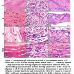
Histopathological Examinations of Kidney Tissue

Renal parenchyma and stroma of normal rats (G1) were normal with keeping features of the nephron units, collecting tubules, papillary and pelvic structures (Figure 7). But renal serial sections of STZ-induced in diabetic rats (60 mg STZ/kg BW, Single ip) (G2) revealed that moderate an array of multifocal necrotic areas (coagulative necrosis) with pyknotic or karryoretic nuclei and deep eosinophilic cytoplasm with keeping a ghost of the tubular and glomerular architecture and distinct inflammatory reaction (Figure 8). Additionally, variable degrees of degenerative changes including cloudy swelling, vacuolar and hydropic degeneration were noticed. Besides this, mild dilatation of some distal convoluted tubules and collecting tubules with partial atrophy of their lining epithelium was recorded. Moreover, a few apoptotic cells were seen in the tubular epithelium together with peculiar perivascular edema and mild to moderate congestion of intertubular and glomerular blood vessels and capillaries, sometimes with erythrocytic extravasations (Figure 8).

Figure 7: Photo-micrograph for rat’s kidney G1 Figure 7: Photo-micrograph for rat’s kidney G1

Click here to View Figure

Figure 8: Photo-micrograph of rat’s kidney G2 Figure 8: Photo-micrograph of rat’s kidney G2

Click here to View Figure

Renal lesions of STZ-induced diabetic rats (60 mg STZ/kg BW, single ip) treated with A. marina 400 mg/kg BW (daily oral dose for 6weeks) (G3) were represented by characteristic perivascular edema with vacuolation of the vascular walls. Some of the nephron units (glomeruli and tubules) were apparently normal, but others showed degenerative and early necrotic and apoptotic changes. The renal papillae and pelvis were apparently normal in most parts; however, a few of them showed mild vascular and tubular dilatations of the surrounding tissues, besides focal exfoliative change in the lining epithelium (Figure 9)

Figure 9: Photo-micrograph of rat’s kidney G3, Figure 9: Photo-micrograph of rat’s kidney G3,

Click here to View Figure

Sections from kidneys of non-diabetic rats, treated with A. marina, 400 mg/kg BW (daily oral dose for 6weeks) (G4), revealed apparently normal nephron units including glomeruli and different types of tubules. The renal papillae, pelvis, medullary rays and blood vessels were also apparently normal. However, a few renal tubular epithelia, especially in the cortex, showed mild degenerative changes, mainly hydropic degeneration (Figure 10).

Figure 10: Photo-micrograph of rat’s kidney G4, Figure 10: Photo-micrograph of rat’s kidney G4,

Click here to View Figure

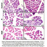
Immunohistochemical Kidney Findings

Kidney sections of normal rats (G1) denoted normal parenchyma free from any apoptotic changes except for a very few cells in the medullary collecting tubules (0.5-1%) /HPF (Figure 11). Meanwhile, renal tissue of STZ-induced diabetic (60 mg STZ/kg BW, Single ip dose) (G2) revealed about 3-5% positive apoptotic reaction of the tubular epithelium, particularly in the medulla (Figure 12). However, kidney sections of STZ-induced diabetic rats (60 mg STZ/kg BW, single ip dose) treated with A. marina 400 mg/kg BW (daily oral dose for 6 weeks) (G3) revealed about 6-8% /HPF of the tubular epithelial cells and 3-5% /HPF of the glomerular cells with positive cytoplasmic apoptotic reaction (Figure 13). Concomitantly, Some of the renal sections of non-diabetic rats treated with A. marina, 400 mg/kg BW (daily oral dose for 6 weeks) (G4) were free of apoptosis, but other sections showed 4-6% cells/HPF of both tubular and glomerular cells with early apoptotic changes as proved by weak brownish cytoplasmic reactivity to caspase-3 (Figure 14).

Figure 11: Photo-micrographs of rat’s kidney Figure 11: Photo-micrographs of rat’s kidney

Click here to View Figure

Figure 12: Photo-micrographs of rat’s kidney, G2, Figure 12: Photo-micrographs of rat’s kidney, G2,

Click here to View Figure

Figure 13: Photo-micrographs of rat’s kidney, G3 Figure 13: Photo-micrographs of rat’s kidney, G3

Click here to View Figure

Figure 14: Photo-micrographs of rat’s kidney, Figure 14: Photo-micrographs of rat’s kidney,G4

Click here to View Figure

Discussion

Diabetes mellitus is a common metabolic disorder, characterized by hypoinsulinemia, hyperglycaemia, hyperlipidemia, hyperaminoacidemia, and disturbances of acute impact on carbohydrates, proteins and lipid metabolisms resulting from serious deleterious defects in either insulin secretion, insulin action, or both.27,28 Remedies developed on the principles of chemical and conventional medications are usually of short affectivity, with a risk of adverse side effects and are usually of high prices, particularly for third world populations. Consequently, treatment of DM with plant derived phytochemicals that are easily obtained and are relatively cheap appears extremely likable.29 Several investigations were applied to review the effectiveness of the plant extracts on diabetes evoked by STZ in many experimental animal tissues.30,31 Many natural products can suppress the enzyme activities responsible for the production of glucose, its absorption, and insulin effectiveness.32,33Other studies have clarified those positively effective components in plant extracts that can modify apoptosis of β-cell and promote insulin action.34-36 The anti-diabetic effects of A. marina have been explored in many studies.37,38

In our current investigation, diabetes was induced by the administration of a single dose of STZ (60 mg/kg B.W), tested animals with blood glucose levels above 250 mg/dl on the 3rd day post treatment of the STZ injection were regarded as diabetic rats.19 In this study, the dramatic elevation of the fasting blood glucose and serum glucose levels (at 6th week) after induction of diabetes were encountered in the diabetic rats as compared with the normal control rats.  The treatment of STZ-induced diabetic rats with A. marina revealed non-significant changes in blood and serum glucose levels in comparison with the diabetic control group. Streptozotocin is a cytotoxic compound especially toxic to the pancreatic β-cells, which are responsible for the production of insulin in mammals. It enters pancreatic β-cells through GLUT2 channels in the cellular plasma membrane, which evidently leads to cellular toxicity and local immune reactions leading finally to hypoinsulinemia and hyperglycemia in animals.39 The harmful effect of STZ on β-cells leads to the development of deficient production of insulin and thus, the elevation of blood glucose level occurs23. Moreover, a significant increase of serum/plasma glucose could provoke additional destruction of β-cells of the pancreas.40

Insulin is the playmaker in diabetes of all types and has a crucial function in glucose homeostasis.   All patients with type-1 diabetes would like to have hormone treatment for good  health (insulin-dependent) unless they receive islet cells or whole organ transplant; several patients with type-2 diabetes could need this hormone once their β-cell function declines over time41. The findings of this study indicated that, at the end of the experimental period, the serum insulin levels were significantly decreased in STZ-induced diabetic rats as compared with the normal control ones. Administration of aqueous extracts of A. marina to diabetic rats resulted in a non- significant change in serum insulin levels when compared with the diabetic control group.

Moreover, creatinine is a well-authenticated as a metabolic derivative of muscle creatine and phosphocreatine and its concentration in serum is proportional to the body muscle mass. Therefore, the amount of creatinine is usually stable and its elevated levels indicate diminished renal function, as it is easily excreted by the nephrons.42 Low serum creatinine levels are positively associated with an increased risk of incident dysglycemia.43,44 Likewise, other studies recorded a positive association between serum creatinine and diabetes prevalence.45 In the current study, there was a significant increase in serum creatinine level of STZ-induced diabetic rats as compared with the normal control rats. The daily administration of aqueous extract of the investigated plants has a no impact on serum creatinine of diabetic animals as compared with STZ-induced diabetic rats. In contrast, Gandomani and Malati46 indicated that A. marina at a dose of 400 mg/kg significantly decreases serum pro-inflammatory cytokines in rats with a consequent reno-protective effect.

It was formally documented that urea is the end product of protein catabolism in the living system and hence, it is synthesized in the liver from ammonia which is produced consequently as a result of the deamination of amino acids. Diabetic nephropathy is one of the major causes of chronic renal failure which is associated with an increased level of BUN and creatinine.45 Another urgent factor is uric acid which is the metabolic end product of purine catabolism and its elevation levels in serum signifies renal functional impairment. Serum uric acid level has been suggested to be associated with the risk of type-2 diabetes. Albeit, biologically, uric acid plays an important role in weakening of insulin resistance in animal models by suppressing the bioavailability of nitric oxide, which is essential for insulin-stimulated glucose uptake.47

In the current study, our evaluation documented a significant increase in a cascade manner embodying increase in serum blood urea nitrogen and uric acid levels of STZ-induced diabetic rats as compared with normal rats. Treatment of STZ-induced diabetic rats with A. marina revealed non-significant change in serum blood urea nitrogen in comparison to the diabetic control group. The results of the current study are contrary to that obtained by Mirazi  et al.48 The authors concluded that hydro-alcoholic extract of A. marina leaves at doses of 400mg or 800mg/kg. B.W could be able to treat renal toxicity induced by CCl4 in male rats with a significant reduction of urea. On the other hand, daily oral administration of A. marina extract to STZ-induced diabetic rats resulted in a significant decrease in serum uric acid levels in comparison to the diabetic control group

Diabetic nephropathy is a major complication of DM and a leading instigator of end-stage renal failure, as a worldwide calamity. The severity of diabetic nephropathy is one of the major factors determining the prognosis of diabetic patients.49 The current investigation on the histo-morphology of the kidney of normal rats showed normal structures of nephron units in almost all the examined sections with keeping features of normal glomeruli, proximal and distal convoluted tubules, loops of Henle, collecting tubules, renal papillae, and renal pelvis. Moreover, the renal blood vessels and intertubular capillaries were in good condition. Renal sections of STZ-induced diabetic rats revealed multifocal coagulative necrotic areas with a pyknotic or karyopyknotic nuclei, deep eosinophilic cytoplasm, and distinct inflammatory reaction. Variable degrees of degenerative changes with mild dilatation of some distal convoluted and collecting tubules along with the partial atrophy of their lining epitheliums were also recorded. Peculiar perivascular edema and mild to moderate congestion of intertubular and glomerular blood vessels and capillaries with erythrocytic extravasations were also seen.

The results of the current study are partially similar to that obtained by Zafar et al.50 who found large aggregates of lymphocytes in the interstitium of rat’s renal tissue following single IP injection of STZ (45 mg/kg B.W). The authors added that an inconstant number of proximal convoluted tubules showed signs of renal-tubular necrosis; their epithelial lining was disrupted and discontinued with pyknotic nuclei, vacuolated cytoplasm, broken cellular membranes and disappearance of brush borders. Nephropathic lesions of the current investigation coincide with that of  Balamash et al.51 This battery of astute scientists have observed that the renal tissue of normal rats showed normal cortex and medulla were intact; renal corpuscles being the main feature of the cortex. Moreover, the outer layer of Bowman’s capsule was lined by simple squamous epithelium and the glomerulus looked like cluster of capillaries covered by the inner layer of Bowman’s capsule. Furthermore, renal tubules in the cortex mainly include the proximal and distal convoluted tubules and these tubules were lined by the cuboidal epithelium.

Kidney lesions in STZ-induced diabetic rats treated with A. marina were represented by characteristic perivascular edema with vacuolation of the vascular walls. Some of the nephron units (glomeruli and tubules) were normal; meanwhile, others showed degenerative and early necrotic changes. The renal papillae and pelvis were normal in most parts; however, a few of them showed mild vascular and tubular dilatations of the surrounding tissue, besides, focal exfoliative changes in the lining epithelium were also seen. With such lesions, a speculated mild decrease in the renal filtration threshold along with a consequent insignificant increase in the creatinine, urea, and uric acids could be expected.

The characteristic renal lesions in the current investigation could lead to induction of dramatic changes in the renal biochemical markers. Such changes could be attributed to the direct nephrotoxic effect of STZ compound and the hyperglycemic secondary toxic metabolites. This information is in agreement with what was revealed earlier that diabetic nephropathy is a major complication of DM and a leading cause of end-stage renal failure worldwide.52 A fact that led to the severity of diabetic nephropathy is one of the major factors determining the prognosis of diabetic patients with the subsequent results that consider it a major determinant of morbidity and mortality in patients with DM. Therefore, previous scientific reports showed that diabetic nephropathy was induced by hyperglycemia via several mechanisms such as oxidative stress along with induction and increase of glycation reaction. Henceforth, the production of highly reactive oxygen radicals was attributed to the hyperglycemic oxidative stress which could lead to prominent cellular cyto-toxicity in different tissues including kidney.53 Moreover, various investigations showed that the level of lipid peroxides as MDA was elevated both in serum and kidney tissues and the levels of the anti-oxidant enzyme were decreased in renal homogenates of diabetic rats.54

Diabetic renal pathologic lesions are contributed to the angio-bio-physiologic complication of DM, which leads to chronic renal problems.55 Apoptosis probably contributes to provoke diabetic nephropathy.56 Apoptotic protein p53 and active caspase-3 are major biomarkers of apoptosis.57 Oxidative stress can activate p53 leading to its nuclear accumulation resulting in cell cycle arrest, apoptosis, or cell removal from the proliferative pool.57 Apoptotic p53 up-regulation is reported in several different tissues in DM, including kidney.59,60 Apoptotic changes were induced by p53 through stimulation of caspases.61 Caspases constitute a group of cysteine proteases which exist in the cell as an inactive zymogen.62 Caspase-3 is the executioner caspase of apoptosis that becomes activated upon cleavage in the apoptotic process leading to the morphological features of apoptosis.63 Elevated levels of active caspase-3 are reported in many renal diseases.64,65

Kidney sections from different experimental groups of the present study revealed 6-8%/HPF in the tubular epithelial cells together with 3-5%/HPF in the glomerular cells of STZ-induced diabetic rats treated with A. marina. The observations of the current study are generally similar to that obtained by Al-Rasheed et al.66 The authors reported that apoptotic markers (BAX and caspase-3) were significantly increased and the anti-apoptotic marker (BCL-2) was decreased in kidneys of STZ-induced diabetic rats. The authors added that topographical localization and double immunohistochemical analysis suggested that podocytes were the main apoptotic cells under these conditions, although endothelial and mesangial cells were also affected. This confirms and extends the previous observations of Susztak et al.56 The authors identified topographically cell type(s) undergoing apoptosis and it was consistent with findings of Meyer et al.67 and Steffes et al.68 where the authors reported that significant podocyte apoptosis could be observed in diabetic patients, parallel with the progression of renal disease. The findings of the present study are in partial agreement with the results obtained by Ahmed et al.69 The authors declared that the pro-inflammatory cytokine TNF-α and the apoptotic mediators (p53 and caspase-3) were remarkably decreased in the kidney of STZ-induced diabetic rats as a result of treatment with VDR activator (paricalcitol), while the expression of anti-apoptotic protein BCL-2 was increased. Furthermore, it should be mentioned consequently that in the aforementioned study, vitamin D was used as a synthetic chemical compound, but it can be naturally obtained from many phytochemical plant sources.70

Finally, the apoptotic p53 protein together with pro-apoptotic proteins were consequently transported into the mitochondria as they encourage an increase in the mitochondrial membrane permeability and lead to energize of cytochrome c, which adheres to the apoptotic protease activating factor 1 (APAF-1) and with the caspase-9 proenzyme, to form a complex called the “apoptosome”. The latter, consecutively, synergized caspase-9 with consequent stimulation of caspase-3 proenzyme to the protease stage, which then sticks to the effector caspase group. Effector caspase-3, -6, and -7 do not need an adaptor protein for dimerization-induced activation. Rather, effector caspases spontaneously dimerize but are only activated upon cleavage between the small and large subunits by an active initiator caspase. Activated effector caspases then cleave a number of protein substrates to initiate apoptosis leading to the subsequent dismantling of cellular components,71-74 evidently the effector caspases induce intracellular protein lysis and the morphological distinctive changes of apoptosis.75 It was reported that apoptosis could aggravate the pathomechanisms of diabetic nephropathy and nephrotoxicity through caspase-3 expression.76 The mitochondrial oncogene product BCL-2, prevented caspase-3 activation during a variety of proapoptotic conditions.77 

Conclusion 

The findings of this study evidently showed that the aqueous leaf extract of A. marina exerted protective effect against streptozotocin-induced nephrotoxicity in male rats as demonstrated by amelioration of the aforementioned biochemical parameters, histopathological and immunohistochemical changes in the kidney sections and restored renal tissue architecture near normal levels. The plant extract showed antihyperglycemic, insulinotropic, and renoprotective actions. Moreover, the findings of this investigation may lead to the development of novel anti-diabetic drugs through the use of up to date technologies which is described as a highly needed imperative

Funding source

There is no funding source

Conflict of Interest

The author of this study did not have any type of conflict of interest.


References

  1. American Diabetes Association, Standards of medical care in diabetes. Clin. Appl. Res. Edu. 2017; 40(1): 1049-5992.
  2. Cerf ME. β-cell dysfunction and insulin resistance. Endocrinol. 2013:4(37): 1-13.
  3. World Health Organization (WHO) and International Diabetes Federation (IDF), 2006. Definition and diagnosis of diabetes mellitus and intermediate hyperglycemia. WHO, Geneva.
  4. Riaz S. Diabetes mellitus. Res. Essays. 2009; 4(5): 367-373.
  5. Macdonald I. a review of recent evidence relating to sugars, insulin resistance, and diabetes. Eur. J. Nutr. 2016; 55(2): 17-23.
  6. Solano M.P., Goldberg R.B. Management of dyslipidemia in diabetes. Rev. 2006; 14(3): 125-135.
  7. Ndisang J.F., Vannacci A., Rastogi S. Insulin resistance, type 1 and type 2 diabetes, and related complications. Diabetes Res. 2017: 1-3.
  8. Kitabchi A.E., Umpierrez G.E., Miles J.M., Fisher J.N. Hyperglycemic crises in adult patients with diabetes. Diabetes Care. 2009; 32(7): 1335–1343.
  9. Chawla A., Chawla R., Jaggi S. Microvascular and macrovascular complications in diabetes mellitus: distinct or continuum? Indian Endocrinol. Metab. 2016; 20(4): 546-551.
  10. Rahmatullah M., Mollik A.H., Azam A.T.M.A., Islam R., Chowdhury A.M., Jahan R., Chowdhury M.H., Rahman T. Ethnobotanical survey of the Santal tribe residing in thakurgaon district, Bangladesh. Eurasian J. Sustain. Agric. 2009; 3(4): 889-898.
  11. Said W.M., Ehsan N.O.M. Morphological and molecular evidence among four heteroforms of Avicennia marina (Forssk) vierh. Am. Sci. 2010; 6(11): 843-856.
  12. Quattrocchi U. CRC World Dictionary of Plant Names: common names, scientific names, eponyms, synonyms, and etymology. 4 vols. CRC Press, Boca Raton, FL. 2000; pp.2896.
  13. Sultana, R.S. Stem and leaf anatomy of Lantana camara-a plant of the Verbenaceae family. Int. J. Curr. Res. Biosci. Plant Biol. 2016; 3(1): 27-31.
  14. Thamizharasan S., Saravanan N.A. Antibacterial potential of mangrove plant Avicennia marina against a clinical pathogen. J. Zool. Stud. 2016; 1(7): 14-16.
  15. Almahasheer H., Duarte C.M., Irigoien X. Phenology and growth dynamics of marina in the central Red Sea. Sci. Rep. 2016; 6: 1-9.
  16. Sahoo,  Mulla N.S., Ansari ZA., Mohandass C. Antibacterial activity of mangrove leaf extracts against human pathogens. Indian J. Pharm. Sci. 2012; 74: 348–351.
  17. Mohamadi J., Havasian M.R. The study of inhibitory effect of aqueous extract leaf of Avicennia marina (Hara) on Candida albicans, In Vitro. J. Pharm. Life Sci. 2017; 8: 5547-5551.
  18. Al-Hariri MT. Comparison the rate of diabetes mellitus induction using streptozotocin dissolved in different solvents in male rats. Comp. Clin. Path. Res. 2012; 1: 96 -99
  19. Gurudeeban S., Kaliamurthi S., Thirugnanasambandam R. Positive regulation of Rhizophora mucronata poir extracts on blood glucose and lipid profile in diabetic rats. Med. 2016; 2: 1-10.
  20. Henry R.J., Cannon D.C., Winkelman J.W. Clinical chemistry: principles and techniques, 11th Ed, Happer and Row Publishers, New York, 1974; pp. 1629.
  21. Young, D.S., 1997. Effects of preanalytical variables on clinical laboratory tests, 2nd Ed. Washington. DC: AACC Press.
  22. Fernández T., Suarez G., Pérez C.L, Acosta T., Clapes S. Influence of diabetes and gestation in blood biochemistry variables in Wistar rats. J. Anim. Sci. 2018; 2: 1-4.
  23. Pournaghi P.,  Sadrkhanlou R.A., Hasanzadeh S., Foroughi A.  An investigation  on body  weights,  blood glucose levels  and  pituitary gonadal  axis  hormones  in  diabetic  and  metformin treated  diabetic  female rats. Res. Forum. 2012; 3: 79-84.
  24. Mazhar F.M., Moawad K.M., Abdel-Gawad M.H. Evidence for a reversing effect of vitamin E or curcumin on some biochemical alterations associated with diabetes in pregnant rats and their fetuses. J. Zool. 2005; 44: 367-388.
  25. Song W., Levin D.S., Varkey J., Post S., Bermudez V.P., Hurwitz J., Tomkinson A.E. A conserved physical and functional interaction between the cell cycle checkpoint clamp loader and DNA ligase I of eukaryotes. Biol. Chem. 2007; 282: 22721-22730
  26. Zar J.H. Biostatistical analysis prentice-hall, eryelwood cliffs. N.J. 1996; pp. 663.
  27. Wadkar K.A., Magdum C.S., Patil S.S., Naikwade NS. Anti-diabetic potential and Indian medicinal plants. Herb. Med. Toxicol. 2008; 2: 45-50.
  28. Al-Attar A.M., Alsalmi FA. Effect of Olea europaealeaves extract on streptozotocin-induced diabetes in male albino rats. Saudi J. Biol. Sci. 2019; 26:118-128.
  29. Bordoloi R., Dutta K.N. A review on herbs used in the treatment of diabetes mellitus. Pharm. Chem. Biol. Sci. 2014; 2: 86-92.
  30. Torrico F., Cepeda M., Guerrero G., Melendez F., Blanco Z., Canelón D.J., Diaz B., Compagnone R.S., Suárez A. Hypoglycaemic effect of Croton cuneatus in streptozotocin-induced diabetic rats. Bras. Farmacogn. 2007; 17: 166-169.
  31. Almalki D,A,, Alghamdi SA. Hepatorenal protective effects of some plant extracts on experimental diabetes in male rats. J. Pharmacol. 2019; 15: 238-247.
  32. Schmidt B., Ribnicky D.M., Poulev A., Logendra S., Cefalu W.T., Raskin I. A natural history of botanical therapeutics. Metabolism 2008; 57: 3-9.
  33. Nazaruk J., Borzym-Kluczyk M. The role of triterpenes in the management of diabetes mellitus and its complications. Rev. 2015; 14: 675-690.
  34. Modak M., Dixit P., Londhe J., Ghaskadbi S., Devasagayam T.P. Indian herbs and herbal drugs used for the treatment of diabetes. Clin. Biochem. Nutr. 2007; 40: 163-173.
  35. Oh Y.S., Jun H.S. Role of bioactive food components in diabetes prevention: effects on beta-cell function and preservation. Metab. Insights. 2014; 6: 51-59.
  36. Saisho Y. β-cell dysfunction: its critical role in prevention andmanagement of type 2 diabetes. World J. Diabetes. 2015; 6: 109-124.
  37. Revathi P., Jeyaseelan S.T, Thirumalaikolundusubramanian P., Prabhu N. An overview of antidiabetic profile of mangrove plants. J. Pharm. Pharm. Sci. 2014; 6: 1-5.
  38. Das S.K., Samantaraya D., Patrab J.K., Samantac L., Thatoid H. Antidiabetic potential of mangrove plants: a review. Front. Life Sci. 2015; 9: 75-88.
  39. Al-Nahdi A.M.T., John A., Raza H. Cytoprotective effects of N-acetylcysteine on streptozotocin-induced oxidative stress and apoptosis in RIN-5F pancreatic β-cells. Cell Physiol. Biochem. 2018; 51: 201-216.
  40. El-Desouki N.I., Tabl G.A., Abdel-Aziz K.K., Salim E.I., Nazeeh N. Improvement in beta-islets of Langerhansin alloxan-induced diabetic rats by erythropoietin and spirulina. Basic. Appl. Zool. 2015; 71: 20-31.
  41. McCulloch D.K, MD., Nathan D.M., MD., Mulder JE., M.D. General Principles of Insulin therapy in Diabetes Mellitus. UpToDate-Online Database. Updated March, 2016. Available at: http://www.uptodate.com/contents/general-principles-of-insulintherapy-in-diabetes-mellitus Accessed December 2017.
  42. Loeb S. Clinical laboratory test: values and implications. Copy right, Springhouse Corporation, Springhouse, Pennsylvania, 1991
  43. Nayak B.S., Butcher D.M., Bujhawan S., Chang D., Chang S., Cabral-Samaroo, Cadan S., Buchoon V., Budhram L., Boyce M., Teelucksingh S. Association of low serum creatinine, abnormal lipid profile, gender, age and ethnicity with type 2 diabetes mellitus in Trinidad and Tobago. Diabetes Res. Clin. Pract. 2011; 91: 342-347.
  44. Takeuchi M., Imano H., Muraki I., Shimizu Y., Hayama-Terada M., Kitamura A., Okada T., Kiyama M., Iso H. Serum creatinine levels and risk of incident type 2 diabetes mellitus or dysglycemia in middle-aged Japanese men: a retrospective cohort study. BMJ Open Diabetes Res. Care. 2018; 6: 1-7.
  45. Chutani A., Pande S. Correlation of serum creatinine and urea with glycemic index and duration of diabetes in type 1 and type 2 diabetes mellitus: a comparative study. J. Physiol. Pharm. Pharmacol. 2017; 7: 914-919.
  46. Gandomani MZ, Malati EF. Evaluation of protective efficacy of Avicennia marina (Forssk.) vierh leaves against complete freund᾽s adjuvant-induced arthritis in wistar. J. Pharm. Res. 2014; 13: 945-951.
  47. Khosla U.M., Zharikov S., Finch J.L., Nakagawa T., Roncal C., Mu W., Krotova K., Block E.R., Prabhakar S., Johnson R.J. Hyperuricemia induces endothelial dysfunction. Kidney Int. 2005; 67: 1739-1742.
  48. Mirazi N., Movassagh S.N., Rafieian-Kopaei M. The protective effect of hydro-alcoholic extract of mangrove (Avicennia marina) leaves on kidney injury induced by carbon tetrachloride in male rats. J. Nephropathol. 2016; 5: 118-122.
  49. Barton M., Sorokin A. Endothelin and the glomerulus in chronic kidney disease. Nephrol. 2015; 35:156-167.
  50. Zafar M., Naqvi S.N., Ahmed M., Kaimkhani, Z.A. Altered liver morphology and enzymes in streptozotocin-induced diabetic rats. J. Morphol. 2009; 27: 719-725.
  51. Balamash K.S., Alkreathy H.M., Al Gahdali E.H., Khoja S.O., Ahmad, A. Comparative biochemical and histopathological studies on the efficacy of metformin and Virgin olive oil against streptozotocin-induced diabetes in Sprague-Dawley rats. J. Diabetes Res. 2018; 2018: 1-10.
  52. Azarkish F., Hashemi K., Talebi A., Kamalinejad M., Soltani N., Pouladian N. Effect of the administration of Solanum nigrum fruit on prevention of diabetic nephropathy in streptozotocin-induced diabetic rats. Res. 2017;9: 325-332.
  53. Barrero A., Quilez-del-Moral J.F., Herrador M.M., Akssira M., Bennamara A., Akkad S., Aitigri M. Oxygenated diterpenes and other constituents from Moroccan Juniperus phoenicea and Juniperus thurifera Africana. Phytochemistry. 2004; 65: 2507-2515.
  54. Rouse, R.L., Stewart, S.R., Thompson, K.L., Zhang, J. Kidney injury biomarkers in hypertensive, diabetic and nephropathy rat models treated with contrast media. Pathol. 2013; 41: 662-680.
  55. Rheinberger M., Böger C.A. Diabetic nephropathy: new insights into diagnosis, prevention and treatment. Med. Wochenschr. 2014; 139: 704-706.
  56. Susztak K., Raff A.C., Schiffer M., Bottinger E.P. Glucose-induced reactive oxygen species cause apoptosis of podocytes and podocyte depletion at the onset of diabetic nephropathy. Diabetes. 2006; 55: 225-233.
  57. Erekat N.S. Cerebellar Purkinje cells die by apoptosis in the shaker mutant rat. Brain Res. 2017; 1657: 323-332.
  58. Horn H.F., Vousden K.H. Coping with stress: multiple ways to activate p53. Oncogene 2007:26:1306-1316.
  59. Qiu L.Q., Sinniah R., Hsu S.I. Coupled induction of iNOS and p53 upregulation in renal resident cells may be linked with apoptotic activity in the pathogenesis of progressive IgA nephropathy. Am. Soc. Nephrol. 2004:15: 2066-2078.
  60. Nakamura H., Matoba S., Iwai-Kanai E., Kimata M., Hoshino A., Nakaoka M, Katamura M., Okawa Y., Ariyoshi M., Mita Y., Ikeda K., Okigaki M., Adachi S., Tanaka H., Takamatsu T., Matsubara H. P53 promotes cardiac dysfunction in diabetic mellitus caused by excessive mitochondrial respiration-mediated reactive oxygen species generation and lipid accumulation. Heart Fail. 2012; 5: 106-115.
  61. Deshpande S.D., Putta S., Wang M., Lai J.Y., Bitzer M., Nelson R.G., Lanting L.L., Kato M., Natarajan R. Transforming growth factor-β-induced cross talk between p53 and a microRNA in the pathogenesis of diabetic nephropathy. Diabetes 2013; 62: 3151-3162.
  62. Fan T.J., Han L.H., Cong R.S., Liang J. Caspase family proteases and apoptosis. Acta Biochim. Biophys. 2005; 37: 719-727.
  63. Erekat N.S. Active caspase-3 upregulation is augmented in at-risk cerebellar  Purkinje cells following inferior olive chemoablation in the shaker mutant rat: an immunofluorescence study. Res. 2019; 41: 234-241.
  64. Bamri-Ezzine S., Ao Z.J., Londoño I., Gingras D., Bendayan M. Apoptosis of tubular epithelial cells in glycogen nephrosis during diabetes. Invest. 2003:83: 1069-1080.
  65. Yano T., Itoh Y., Matsuo M., Kawashiri T., Egashira N., Oishi R. Involvement of both tumor necrosis factor-alpha-induced necrosis and p53-mediated caspase-dependent apoptosis in nephrotoxicity of cisplatin. 2007; 12: 1901-1909.
  66. Al-Rasheed N.M., Al-Rasheed N.M., Bassiouni Y.A., Hasan I.H.,  Al-Amin M.A., Al-Ajmi H.N., Mahmoud A.M. Simvastatin ameliorates diabetic nephropathy by attenuating oxidative stress and apoptosis in a rat model of STZ-induced type 1 diabetes. Pharmacother. 2018; 105: 290-298.
  67. Meyer T.W., Bennett P.H., Nelson R.G. Podocyte number predicts long-term urinary albumin excretion in Pima Indians with type II diabetes and microalbuminuria. 1999; 42: 1341-1344.
  68. Steffes M.W., Schmidt D., McCrery R., Basgen J.M. Glomerular cell number in normal subjects and in type 1 diabetic patients. Kidney Int. 2001; 59: 2104-2113.
  69. Ahmed O.M., Ali T.M., Abdel Gaid M.A., Elberry A. Effects of enalapril and paricalcitol treatment on diabetic nephropathy and renal expressions of TNF-α, P53, caspase-3 and Bcl-2 in STZ-induced diabetic rats. bioRxiv 2019;2019: 1-33.
  70. JäpeltB., Jakobsen J.  Vitamin D in plants: a review of occurrence, analysis, and biosynthesis. Front. Plant Sci. 2013; 4: 1-20.
  71. Fischer H., Koenig U., Eckhart L., Tschachler E. Human caspase 12 has acquired deleterious mutations. Biochem. Biophys. Res. Commun. 2002; 293: 722726.
  72. Lüthi A.U., Martin S.J. The CASBAH: a searchable database of caspase substrates. Cell Death Differ. 2007; 14: 641-650.
  73. Taylor R.C., Cullen S.P., Martin S.J. Apoptosis: controlled demolition at the cellular level. Rev. Mol. Cell Biol. 2008; 9: 231-241.
  74. Kurokawa M., Kornbluth S. Caspases and kinases in a death grip. Cell. 2009; 138: 838-854.
  75. Sznarkowska A., Olszewski R., Zawacka-Pankau J. Pharmacological activation of tumor suppressor, wild-type p53 as a promising strategy to fight cancer. Postepy Hig. Med. Dosw.  2010;64: 396-407
  76. El-Beshbishy H.A., Bahashwan S.A., Aly H.A., Fakher H.A. Abrogation of cisplatin-induced nephrotoxicity in mice by alpha lipoic acid through ameliorating oxidative stress and enhancing gene expression of antioxidant enzymes. J. Pharmacol. 2011; 668: 278-284.
  77. Zhang S., Ong C.N., Shen H.M. Involvement of proapoptotic Bcl-2 family members in parthenolide-induced mitochondrial dysfunction and apoptosis. Cancer Lett. 2004; 211: 175-188.

Malted Barley Improved the Structure and Function of Gastrocnemius Muscle of Hypercholesterolemic Mother Rats and their Offspring

$
0
0

Introduction

The skeletal muscles make up about 55% of human body mass and play important roles in contraction and relaxation. They are the main site for glucose utilization, oxidation of fatty acids and metabolism of proteins (Kuo and Ehrlich, 2015).

Skeletal muscles are also made up of muscle fibers population (I, IIA, IIX and IIB). They are regulated by their myosin heavy chains, which control the contraction and relaxation (Hoh, 2011). This involved the conversion of the chemical energy through hydrolysis of ATP into contraction of the cytoskeletal apparatus (Schiaffino and Reggiani, 2011; Pataky et al., 2019). Two points are important of the muscle. First of it is the higher inclusion of cholesterol in their sarcolemma which facilitate the membrane fluidity (Barrientos et al., 2017). The second its high content of mitochondria required for ATP production.The muscle fibers were highly susceptible to  ROS-induced damage of their fibers (Zhang et al., 2017).

Myocytes are highly susceptible to the obesity associated inflammation and generation of ROS through the reduction of  superoxide dismutase, catalase and glutathione peroxidase (Moylan and Reid, 2007).  Consumption of fat diet was found to decrease the actin filaments (F-actin) necessary to control glucose transporter 4 (GLUT4) and predicting early insulin resistance in the skeletal muscle (Grice et al., 2019). Oxidative stress associated with obesity is significantly involved in establishing insulin resistance in both adipocytes and myocytes (Gozalez-Franquesa and Patti, 2017). High fat diet significantly decreased type I fiber In lumbar muscles, while increased type II. Such phenomena can indicate skeletal muscle reaction to overload of dietary lipid to regulate metabolic homeostasis (Hua et al., 2017).

Soaked barley foods are a prebiotic that increased the production luminal butyrate. It provided the enzymes (α-amylase, β-amylase) required for various forms of sugar, such as monosaccharide glucose, disaccharide maltose , disaccharide maltotriose, and maltodextrines (Hanai et al., 2004; Kranz et al., 2015). Germinating barley food attenuated inflammation by reducing sera levels of tumor necrosis factor-α and IL-6 and 8  of ulcerative colitis (Faghfoori et al., 2011) modulating gut microbiota (Zhong et al.2015), and anti-hyperglycaemia (Ramakrishna et al., 2017).

Little of work is available about the phytomedicinal importance of fermented barley against the ingestion of hypercholesterolemic diet which induces skeletal muscle damage. The present work focused on assessing the histopathological, ultrastructural and biochemical changes of high cholesterol diet on gastrocnemius muscle of mother rats and their offspring as well as the effect of ingested soaked barley grain.

Material and Methods

Ethical Consideration

This research was carried out according to the National Institute of Health, s guide to the use of the laboratory animals and supported by the Mansoura University,s Egyptian Committee for Animal Care and Bioethics. 

Preparation of a High Cholesterol Diet and Assessment of Hyperlipidemia

It was done by ingestion of 3% cholesterol diet containing 10% animal fat, 2% cholic acid and 1% thiouracil in addition to the normal diet for 4 months prior to conception and throughout pregnancy and lactation period (Enkhmaa  et al., 2005). For critical assessment of hyperlipidemia, under sterile condition, the blood is aspirated from the ocular artery by capillary tube, centrifuged at 2000 rpm and sera were removed. Total Cholesterol (Deeg and Ziegenhorn, 1983) and LDL (Friedewald et al., 1972) were estimated.

Diets Containing Soaked Barley

The standard diet is composed of 20% malted barley. The hypercholesterolemic groups were fed on diet containing 20% barley prior to conception and during the gestation and lactation period.

Experimental Animal

Sixty virgin female and twenty adult male albino rats (Rattus norvegicus) weighing approximately 100gm body weight, obtained from Helwan Breading Farm, Ministry of Health, Egypt, have been used for experimental investigation. It has been provided free access to food and water ad libitum. They kept in good ventilation with 12 hour light/dark cycle. Virgin females were allowed for mating with fertile male (3 female/1male) for overnight. In the next morning, visualizing sperm in the vaginal smear determine the onset of gestation. The pregnant rats were arranged into 4 groups (n=15) including; control (C), barley supplemented group (B), hypercholesterolemic-group (H) and hypercholesterolemic & soaked barley group (H+B. Mothers of the studied groups were anaesthetized by subcutaneous injection of sodium phenobarbitone (9.1 mg/kg) at 3 weeks post-partum and sacrificed. Blood was collected from the heart of both mothers and their offspring, allowed to coagulate, and sera were separated and kept in refrigerator. At the same time, their gastrocnemius were dissected and investigated as follows:

Histological Investigation

The specimens were fixed in 10 % phosphate buffered formalin (pH 7.4), dehydrated in the highest degree of ethyl alcohol, cleared in toluene and mounted in melted paraplast 58-62ºC. Five µm histological sections were cut, stained with hematoxylin & eosin and examined under a bright field light microscope.

Transmission Electron Microscopy (TEM)

Extra- specimens were fixed in 2.5% in 0.1 M phosphate buffer (pH 7.4), followed by 1 % osmium tetraoxide. They were then dehydrated in ascending ethyl alcohol, cleared with acetone and embeded in epoxy resin. Ultrathin sections were cut on a LKB Ultratome IV (LKB Instruments, Bromma, Sweden) with a diamond knife and mounted on grids, stained with uranyl acetate and lead citrate, and viewed under a Joel 100CX electron transmission microscope (Musashino 3-chome, Akishima, Tokyo 196-8558, Japan).

Biochemical Assessments of Sera Lipid Profiles

Sera levels of total cholesterol (TC) (Deeg and Ziegenhorn, 1983), Triglycerides (TG) (Fossati and Prencipe, 1982) and high density lipoproteins (HDL) (Grove, 1979) were assayed. Low density lipoproteins (LDL) was calculated from the total concentrations of cholesterol (TC), HDL-cholesterol and triglycerides according to Friedewald et al (1972). The glucose was determined by blood glucometers one touch ultra (Life Scan Milipitas, CA, USA).

Assessment of Sera Antioxidants Glutathione-S-Transferase and Superoxide Dismutase Activities

Glutathione S-transferase (GST) is determined through the conjugation of 1- chloro- 2,4- dinitrobenzene with reduced glutathione followed by measuring the absorbance at 340 nm ( Prins and loose, 1969). Also, determination of superoxide dismutase (SOD) depends on the reduction of nitroblue tetrazolium (NBT) by superoxide radicals to the blue colored formazan and assayed calorimetrically at 560 nm (Nishikimi et al. (1972),

Determination of Serum MDA Content

It is evaluated on the basis of red colour development according to Ohkawa et al. (1979), and calculated at 532 nm for specifying the degree of peroxidation and expressed as nmol/ mg protein.

Determination of Serum Caspase-3

It is determined calorimetrically by using a Stressgen kit (catalog No. 907-013). The cleavage of the peptide can be quantitated spectrophotometrically at a wavelength of 405nm. The level of caspase enzymatic activity in the cell lysate is directly proportional to the color reaction.

Determination of Muscle Actin Beta and Keratin 18

Actin beta (ACTb, catalogue no. SEB340Mi) and keratin 18 (KRT18, catalogue no. SEB231Ra) were assayed by using Enzyme-linked Immunosorbent Assay Kit of Cloud-Clone Corp.

Assessment of Muscle Isoenzymes Electrophoresis

Fresh samples of gastrocnemius muscle of the studied groups were homogenized and analyzed by SDS-PAGE (Laemeli, 1970).The protein content was separated and determined (Lowry et al., 1951). Electrophoresis was done at 4°C in polyacrylamide gel and protein bands were visualized by staining with coomassie blue R-250 (60 mg/L) in an acidic medium (Andrew, 1970). The assayed enzymes are:

Glucose-6-Phosphate Dehydrogenase (G6PD)

The electrophoretic buffer contained 5mM tris, 80 mM aspartate and 20 µM NADP+ at (pH 7.4) and visualized the enzyme activity at 30°C in a media composed of  in 20 mL, 1.2 mM tris-phosphate buffer (pH 8.5), 25% (v/v) glycerol, 30 µM glucose-6-phosphate, 4 mM NADP+, 6mg p-nitroblue tetrazolium and 0.5mg phenazine methosulfate (Gaal et al., 1980).

Lactic Dehydrogenase Isoenzymes

After electrophoresis, the gels were incubated into media containing  18.4 mL H2O, 4mL 1M tris, 12mL tetrazolium-blue, 4 mL phenazine methosulphate, 4mL Na-lactate  and 1.3 mL NAD  to develop color reaction for 20 min. Investigating the gel, the appearance or absence of a certain isoenzymatic band of the isoenzymes were  recorded (to  Lehnert and Berlet,1979).

Statistical Analysis

Statistics were calculated with SPSS for windows version 15.0 (SPSS Inc., Chicago, IL, USA). The means value obtained in the different groups were compared by one-way post-hoc analysis of variance (ANOVA) test. All results were expressed as mean±standard error (SE) and significance were defined at p <0.05 and highly significant at p<0.01.

Results

Light and Ultra-Structural Observations

Gastrocnemius of Mother Rats

At light microscopic level, mother rats ingested a high cholesterol diet showed a considerable widening of the interstitial spaces between the muscle fibers. The muscle fibers possessed focal necrosis (Fig. 1A2). On the other side, barley supplementation in diet containing a high fat diet showed restoration of the muscle fibers. Its histological picture seemed to be of nearly normal characteristic feature (Fig.1A3). Regard the control (Fig.1 A) and barley fed groups (Fig.1 A1)

Figure 1: Photomicrograph of horizontal section of gastrocnemius muscle. Figure 1: Photomicrograph of horizontal section of gastrocnemius muscle.

Click here to View Figure

At transmission electron microscopy, compared to the control (Fig.3 A), the myofibrils were disorganized and lacked normally oriented muscle bands in mother fed on a high cholesterol diet. The nuclei of the myocytes become pyknotic. They were enclosed by electron dense heterochromatin. Many necrotic foci were diffusely scattered in the muscle fibers. Lipid deposits were appeared in between the myofibers. Damaging of the mitochondria was become evident (Fig. 3 A1 and A2).

In mother rat ingested barley and a hypercholesterolemic diet, the muscle fibers restored their regularly orientated muscle bands. Glycogen granules and mitochondria were detected in between muscle fibrils (Fig. 3A-A3).

Gastrocnemius of Offspring

At light microscopy level, offspring of mother ingested a high cholesterol diet showed abnormal structure of muscle fibers characterized by expansion of the endomysium and increased perimysial connective tissue. The thickening of muscle fibers varied markedly between each other. Some of the muscle fibers appeared damaging and fragmented (Fig.1B2 & C2). In cross sections, the muscle fibers become widely separated and possessed dense infiltration of leukocytes. In 1 week -old, there was a remarked indistinct cross striation associated with the presence of necrotic areas. Dense inflammation associated with necrotic foci and lipid deposits were observed in 3-week-old offspring (Fig. 2 B1, B2). Regard the normal structural of control and barley fed group (Figs. 1 B and C, 2 A1 and A2).

Figure 2: Photomicrograph of cross section of gastrocnemius muscle of offspring. Figure 2: Photomicrograph of cross section of gastrocnemius muscle of offspring.

Click here to View Figure

In offspring of mother’s ingested barley and a high cholesterol diet, the histological picture of the gastrocnemius appeared comparatively improved (Figs. 1 B3, C3 and 2 C1, C2).

At transmission electron microscopy, the muscle of offspring of hypercholesterolemic mother showed a detected disorganization of muscle fibers with corrugated sarcolemma. The myocyte nuclei become pyknotic and enclosed by electron dense chromatin. Their nucleolar envelope become convoluted (Fig.3 B1, B2 and C1, C2) compared to the normal ordinary myocytes of H, A, I and Z bands in control (Fig. 3 B and C).

Figure 3: Transmission electron micrograph of gastrocnemius muscle. Figure 3: Transmission electron micrograph of gastrocnemius muscle.

Click here to View Figure

On the other hand, offspring of mother ingested barley containing a high cholesterol diet preserved most of the elementary structure of the muscle associated with nearly almost the normal muscle bands of the muscle fibers. The distribution of nuclear chromatin appeared to be of normal characteristic structure. The intefibillar structure showed normal mitochondrial distribution with characteristic internal cristae (Figs. 3 B3 and C3).

Biochemical Observations of Mothers

Compared to both of the control and barley fed mother rats, sera of mothers ingested a high cholesterol diet showed significant decrease of the activities of both glutathione s-transferase  and superoxide dismutase with a concomitant significantly increase of MDA and casp3. However, there was a detected decrease of MDA and casp 3 and increase to almost normal value of the assayed antioxidant enzymes in muscle of hypercholesterolemic mothers ingested barley (Figs. 4 and 5).

Figure 4: Diagram representing sera level of GST (A) and SOD (B) Figure 4: Diagram representing sera level of GST (A) and SOD (B)

Click here to View Figure

Figure 5: Diagram representing sera levels of MDA (A) and caspas3 (B) Figure 5: Diagram representing sera levels of MDA (A) and caspas3 (B)

Click here to View Figure

In experimental mother rats fed on a high cholesterol diet, there was a detected significant decrease of the sera total protein and B-actin s with a concomitant increase of keratin. However, ingestion of barley and a high cholesterol diet, showed a detected improvement of the assayed parameters but their levels were still less than the control values (Figs. 6& 7).

Figure 6: Diagram representing gastrocnemius muscle total protein of mother rats Figure 6: Diagram representing gastrocnemius muscle total protein of mother rats

Click here to View Figure

Figure 7: Diagram representing gastrocnemius muscle Figure 7: Diagram representing gastrocnemius muscle

Click here to View Figure

Biochemical Observations of Offspring

Offspring of mothers ingested a high cholesterol diet possessed a significant depletion of the activities of both the sera  GST and SOD (Fig.4 A and B). The decrease of the antioxidant enzymes were associated with increased sera levels of MDA and Casp-3. The mentioned dramatic alterations were ameliorated in those of mothers ingested barley plus a high cholesterol diet but were still not matched with the control values (Fig. 5 A and B).

In one and three week-old offspring of mothers ingested a high cholesterol diet, the muscle contents of total protein and B-actin were markedly decreased coincides with a marked increase of keratin. However, in those of mothers ingested  barley plus a high cholesterol diet, the muscle contents of the assayed actin and protein were significantly improved but still differed from the control values (Figs. 6 & 7 A and B).

Sodium Dodecylsulfate–Polyacrylamide Gel Electrophoresis (SDS-PAGE)

Sodium dodecylsulfate–polyacrylamide gel electrophoresis of gastrocnemius muscle of mother rat ingested a high cholesterol diet and their offspring showed a decreased expression of the protein bands compared to the control and barley fed group (Fig. 8).

Figure 8: Diagram representing SDS-PAGE protein expression of gastrocnemius muscle Figure 8: Diagram representing SDS-PAGE protein expression of gastrocnemius muscle

Click here to View Figure

Lactic Dehydrogenase Isoenzymes Electrophoresis (LDH)

Following differential LDH isoenzymes of gastrocnemius muscle of mother, LDH expressed five isoenzyme fractions. In mother ingested a high cholesterol diet, the second and third isoenzyme fractions become denser, while the fractions IV and V appeared faintly compared to the other groups. On the other side, 1week-old offspring showed increased densities of the isoenzyme fractions II, III, IV and V compared to the other groups. The densities of the isoenzyme fractions were comparatively decreased in 3week-old offspring, compared to that of the control and barley fed groups. On offspring of mother ingested a high cholesterol diet containing barley, the LDH isoenzymes of the gastrocnemius muscle were improved (Fig. 9).

Figure 9: Diagram representing LDH and G6-PHD isoenzyme electrophoresis Figure 9: Diagram representing LDH and G6-PHD isoenzyme electrophoresis

Click here to View Figure

Glucose-6-Phosphatase Dehydrogenase Isoenzyme Electrophoresis (G6PD)

In mother G6-PHD, three isoenzyme fractions were identified in gastrocnemius muscle. Mother ingested a high cholesterol diet showed faint expression of the isoenzyme fractions I, II & III comparing with either control or barley group. In mother ingested a high cholesterol diet containing barley, there was a detected newly isoenzyme fraction IV and faint expression of fraction III.

In 1week-old offspring G6-PDH, four isoenzyme fractions are observed. Offspring of mother rats ingested on a high cholesterol diet showed a weak expression of the isoenzyme fractions III and IV compared to either the control or barley groups. In offspring of mother ingested a high cholesterol diet containing barley, there was a detected decreased expression of the isoenzyme fraction III and newly expression of the isoenzyme fraction V. In 3week-old offspring G6-PDH, four isoenzyme fractions were observed. Offspring of mothers ingested on a high cholesterol diet showed decreased expression of the isoenzyme fractions compared to the control or barley group. In those maternally fed on a high cholesterol diet containing barley, decreased expression of the isoenzyme fractions were observed compared to the other groups (Fig. 9).

Discussion

The present study showed that mothers ingested a high cholesterol diet was abnormally disorganized the gastrocnemius muscle clarified by expanding of the interstitial space, focal necrosis and disorganized muscle bands. The affected myocytes had compacted nuclear chromatin materials that displayed sign of pyknosis. Leukocytic infiltration was detected in the necrotic zone.  Ultrastructurally, lipid deposits were observed in between the muscle fibers. The alterations in mother rats reflected the abnormal structural organization of gastrocnemius muscle in their offspring. The muscle fibers varied in thickness, expanding of the endomysium, increased perimysial connective tissue, splitting and damaged myofibrils as well as dense collection of inflammatory cells especially in necrotic patches.

The present results are consistent with authors who reported increased accumulation of lipid in  the soleus and tibialis anterior muscles  associated damage of muscle fibers (Collino et al., 2014) and atrophy of  the tibialis anterior of mice ingested a high fat diet (Abrigo et al., 2016). Also, mitochondrial damage and increased intracellular oxidative stress were reported in skeletal muscle in obese diabetic patients  (Dos Santos et al., 2018).

Rats fed  on either  45 percent  or 60 percent fat diet  for 3 weeks  had injured   gastrocnemius assessed by  acidophilic staining and deposition of lipid droplets in between muscle fibers (Ickin et al., 2015). Extra-myocellular deposition of lipid coincides with decrease of type 1 fiber and increase of type 2 in lumbar muscle (Hua et al., 2017).

The observed increase of sera maternal levels of LDL, and cholesterol associated depletion of the antioxidant enzyme defense of glutathione s-transferase and superoxide dismutase as well as increased lipid peroxidation assessed by malondialdhyde reflected the increased oxidative stress and muscle damage. This was represented by increased expression of sera levels of caspase 3.  The depletion of the antioxidant enzymes are known to increase the production of reactive oxygen species superoxide, post– myocyte damage resulting from leakage electron from the mitochondrial transport chain (Kim et al., 2008).

Also, the diseased gastrocnemius muscle of mother rats ingested a high cholesterol diet and their offspring was explained decreased protein synthesis and missing of protein bands following SDS-PAGE analysis, associated increased expression of LDH isoenzyme fractions predicting the muscle damage. At the same time, the G6-PD isoenzymes expression was decreased. Glucose-6-phosphate dehydrogenase  is known to be involved in the myosin light chain phosphorylation via pentose phosphate pathway which is a major source of NADPH, which regulates various enzymatic (Gupta et al., 2011). This can explain the muscle damage.

In addition, the gastrocnemius muscle of mother rats ingested a high cholesterol diet and their offspring showed a significant decrease of total protein and B-actin concomitant with increased keratin content. This finding is consistent with that of  Lóry et al. (2019) who reported depletion of protein in muscle of obese rat due to reduction of the aminopeptidase A activity and angiotensin-converting enzyme in the skeletal muscle leading to dysfunction of myosin heavy chain.

Also, the high cholesterol diet was found to decrease the protein muscle content and β-actin correlates with increased muscle keratin. β-actin is known to be localized in stress fibers of circular bundles. This may be resulted from decreased phosphorylation (Sun et al., 2019) and associated depletion of cortical filamentous actin of glucose transporter 4 (Grice et al., 2019).  Reduction of protein and β-actin explained the muscle dysfunction.

Keratin is also the intermediate protein filament required for transducing contractile force (Muried et al., 2020). Overexpression of keratin is associated with the development of muscular atrophy Toivola et al., 2008).

On the other side, barley supplementation to mother  rat ingested a high cholesterol diet restored the striation of muscle fibers associated with increased the assayed antioxidant enzymes and decreased the MDA marker of oxidative stress and caspase 3 that assessed cell death. This improvement of muscular structure reflected the increase of G6PD and protenin and β-catenin content.

Barley contain varieties of nutrients such as β-glucan which decrease blood cholesterol   (Aludatt et al., 2012) in animals and human (Wilson et al., 2004; AbuMweis et al., 2010). Also, it is rich in antioxidant and phenolic components (Wilson et al., 2004; AbuMweis et al.,2010), which  increased the high-density lipoprotein (HDL) (Shimizu et al.,2019), that scavenge free radicals in cell membranes, preventing damage to their cellular lipids and proteins or DNA (Lobo et al.,2010)

The authors concluded that supplementation with barley has therapeutic potential against muscular dystrophy caused by hypercholesterolemia. The dietary soaked barley restored almost the antioxidant enzymes and reduced oxidative stress. The gastrocnemius muscle restored almost the normal structural pattern and protein and actin content for performing its function in both mother rats and their offspring.

Conflict of Interest

The authors declare that there is no conflict of interest.

References

  1. Abrigo J, Rivera JC, Aravena J et al. High Fat Diet-induced skeletal muscle wasting is decreased by mesenchymal stem stem cells administration: Implications on oxidative stress, Ubiquitin proteasome pathway activation, and myonuclear apoptosis. Oxid Med Cell Longev. 2016; 2016 (9047821), 13 pages.
  2. AbuMweis SS, Jew S, Ames NP. β-glucan from barley and its lipid-lowering capacity: a meta-analysis of randomized, controlled trials. Eur J Clin Nutr. 2010; 64(12):1472-1480.
  3. Aludatt MH, Abu-Zaiton A, Alrababah M et al. Anti-oxidant, Anti-diabetic, and anti-hypertensive eEffects of extracted phenolics and hydrolyzed peptides from barley protein eractions. Int J. Food Prop. 2012; 15(4): 781-795.
  4. Andrews AT. Electrophoresis: theory, techniques, biochemical and clinical applications, clarendon press, Oxford.1986
  5. Andrich DE, Melbouci L, Ou Y et al. A Short-term high-fat diet alters glutathione levels and IL-6 gene expression in oxidative skeletal muscles of young rats. Front Physiol. 2019; 10: 372.
  6. Barrientos G,Sánchez-Aguilera P, Jaimovich E et al. Membrane cholesterol in skeletal muscle: A novel player in excitation-contraction coupling and insulin resistance. J Diabetes Res. 2017; 2017: 3941898.
  7. Bartłomiej S, Justyna RK, Ewa N. Bioactive compounds in cereal grains – occurrence, structure, technological significance and nutritional benefits – a review. Food Sci Technol Int. 2012; 18(6): 559-568.
  8. Collino M, Mastrocola R, Nigro D et al. Variability in myosteatosis and insulin resistance induced by high- fat diet in mouse skeletal muscles. Biomed Res Int. 2014; 2014:569623.
  9. Deeg R, Ziegenhorn J. Kinetic enzymic method for automated determination of total cholesterol in serum. 1983; 29: 1798–1802.
  10. Dos Santos JM, de Oliveira DS, Moreli ML et al. The role of mitochondrial DNA damage at skeletal muscle oxidative stress on the development of type 2 diabetes. Mol Cell Biochem. 2018;449(1-2):251-255.
  11. Enkhmaa B, Shiwaku K, Anuurad E et al. Prevalence of the metabolic syndrome using the Third Report of the National Cholesterol Educational Program Expert Panel on Detection, Evaluation, and treatment of high blood cholesterol in adults (ATP III) and the modified ATP III definitions for Japanese and Mongolians. Clin Chim Acta. 2005; 352(1-2):105-113.
  12. Faghfoori Z, Navai L, Shakerhosseini R et al. Effects of an oral supplementation of germinated barley foodstuff on serum tumor necrosis factor-alpha, interleukin-6 and -8 in patients with ulcerative colitis. Ann Clin Biochem.2011; 48(Pt 3):233-7.
  13. Fossati P, Prencipe L . Serum triglycerides determined colorimetrically with an enzyme that proceduces hydrogen peroxide. Chem. 1982; 28: 2077–2080.
  14. Friedewald WT, Levy RI, Fredrickson DS. Estimation of low density lipoprotein cholesterol in plasma without use preparative ultracentrifuge. Chem. 1972; 18: 499–502.
  15. Gaal O, Medgyesi GA, Vereczkey L. Electrophoresis in the separation of biological macromolecules O. Gaal, G. A. Medgyesi, L. Vereczkey John Wiley & Sons (1980) Price-$57.75. J Immunopharmacol.. 1980; 2 (4): 573-574.
  16. Gamel T, Abdel-Aal ESM. Phenolic acids and antioxidant properties of barley wholegrain and pearling fractions. Food Sci. 2012; 21(2):118-131.
  17. Gonzalez-Franquesa A, Patti ME. Insulin resistance and mitochondrial dysfunction. Adv Exp Med Biol. 2017; 982:465–520.
  18. Grice BA, Barton KJ, Covert JD et al. Excess membrane cholesterol is an early contributing reversible aspect of skeletal muscle insulin resistance in C57BL/6NJ mice fed a Western-style high-fat diet. Am J Physiol Endocrinol Metab. 2019;317(2):E362-E373.
  19. Grove TH. Effect of reagent PH on determination of the high-density lipoprotein cholesterol by precipitation with sodium phototungestate-magnesium. Chem. 1979; 25: 560–564.
  20. Gupte RS, Ata H, Rawat D et al. Glucose-6-phosphate dehydrogenase is a regulator of vascular smooth muscle contraction. Antioxid Redox Signal. 2011 ;14(4):543-558.
  21. Hanai H, Kanauchi O, Mitsuyama K et al. Germinated barley food-stuff prolongs remission in patients with ulcerative colitis. Int J Mol Med. 2004;13(5):643-647.
  22. Hoh JF. Muscle fiber types and function. Curr Opin Rheumatol. 1992; 4(6):801–808.
  23. Hua N, Takahashi H, Yee GM et al. Influence of muscle fiber type composition on early fat accumulation under high-fat diet challenge. PLoS One. 2017;12(8): e0182430.
  24. Ickin Gulen M, Guven Bagla A et al. Histopathological changes in rat pancreas and skeletal muscle associated with high fat diet induced insulin resistance. Biotech Histochem. 2015; 90(7):495-505.
  25. Kim JA, Wei Y, Sowers JR. Role of mitochondrial dysfunction in insulin resistance. Circ Res. 2008; 102: 401–414.
  26. Kranz B, Koch M, Schapfl M et al. Investigation of the germination of barley and wheat grains with a design of experiments for the production of hydrolases. Food Technol Biotechnol. 2015; 53(2):127–135.
  27. Kuo IY, Ehrlich BE. Signaling in muscle contraction. Cold Spring Harb Perspect Biol. 2015 ;7(2):a006023
  28. Laemmli UK. Cleavage of structural proteins during the assembly of the head of the bacteriophage T4. Nature. 1970; 227:680-685.
  29. Lehnert T, Berlet HH. Selective inactivation of lactate dehydrogenase of rat tissues by sodium deoxycholate. Biochem J.1979; 177:813–8.
  30. Lobo V, Patil A, Phatak A et al. Free radicals, antioxidants and functional foods: Impact on human health. Pharmacogn Rev. 2010; 4(8): 118-126.
  31. Lowry OH, Rosebrough NJ, Farr AL, Randall RJ. Protein measurement with the Folin phenol reagent. J Biol Chem. 1951; 193(1): 265-275.
  32. Moylan JS, Reid MB. Oxidative stress, chronic disease, and muscle wasting. Muscle Nerve. 2007;35(4):411–429.
  33. Muriel JM, O’Neill A, Kerr JP et al. Keratin 18 is an integral part of the intermediate filament network in murine skeletal muscle. Am J Physiol Cell Physiol. 2020;318(1):C215–C224.
  34. Nishikimi M, Appaji N, Yagi K. The occurrence of superoxide anion in the reaction of reduced phenazine methosulfate and molecular oxygen. Biochem Biophys Res Commun. 1972; 46(2), 849-854.
  35. Ohkawa H, Ohishi N, Yagi K. Assay for lipid peroxides in animal tissues by thiobarbituric acid reaction. Anal Biochem. 1979; 95(2): 351-358.
  36. Pataky MW, Yu CS, Nie Y et al. Skeletal muscle fiber type-selective effects of acute exercise on insulin-stimulated glucose uptake in insulin-resistant, high-fat-fed rats. Am J Physiol Endocrinol Metab. 2019; 316(5): E695–E706.
  37. Pinho RA, Sepa-Kishi DM, Bikopoulos G et al. High-fat diet induces skeletal muscle oxidative stress in a fiber type-dependent manner in rats. Free Radic Biol Med. 2017;110:381–389.
  38. Prins HK, Loose JA . Glutathione “chapter 4” in biochemical methods in red cell genetics. Edited by J.J. Yunis. Academic press, N.Y.D. London. 1969, 126-129.
  39. Ramakrishna R, Sarkar D, Manduri A et al. Shetty K. Improving phenolic bioactive-linked anti-hyperglycemic functions of dark germinated barley sprouts (Hordeum vulgare L.) using seed elicitation strategy. J Food Sci Technol. 2017; 54(11):3666–3678.
  40. Schiaffino S, Reggiani C. Fiber types in mammalian skeletal muscles. Physiol Rev. 2011; 91(4):1447–1531.
  41. Shimizu C, Wakita Y, Kihara M et al. Association of lifelong intake of barley diet with healthy aging: Changes in physical and cognitive functions and intestinal microbiome in senescence-accelerated mouse-prone 8 (SAMP8). Nutrients 2019; 11, 1770.
  42. Sun YN, Huang JQ, Chen ZZ et al. Amyotrophy induced by a high-fat diet is closely related to inflammation and protein degradation determined by quantitative phosphoproteomic analysis in skeletal muscle of C57BL/6 J Mice. J Nutr. 2020; 150(2):294-302.
  43. Toivola DM, Nakamichi I, Strnad P et al. Keratin overexpression levels correlate with the content of spontaneous pancreatic injury. Am J Pathol. 2008; 172(4):882-892.
  44. Wilson TA, Nicolosi RJ, Delaney B et al. Reduced and high molecular weight barley beta-glucans decrease plasma total and non-HDL-cholesterol in hypercholesterolemic Syrian golden hamsters. J Nutr. 2004; 134(10): 2617-2622.
  45. Zhang L, Zhou Y, Wu W et al. Skeletal muscle-specific overexpression of PGC-1α induces fiber-type conversion through enhanced mitochondrial respiration and fatty acid oxidation in mice and pigs. Int J Biol Sci. 2017;13(9):1152–1162.
  46. Zheng XK, Zhang L, Wang WW et al. Anti-diabetic activity and potential mechanism of total flavonoids of Selaginella tamariscina (Beauv.) Spring in rats induced by high fat diet and low dose STZ. J Ethnopharmacol. 2011;137(1):662–668.
  47. Zhong Y, Nyman M, Fåk F. Modulation of gut microbiota in rats fed high-fat diets by processing whole-grain barley to barley malt. Mol Nutr Food Res. 2015; 59(10):2066–2076.

Modification of Paraburkholderia TALE-Like Protein to Activate Transcription of Target Genes in Plant

$
0
0

Introduction

Many approaches to controlling gene expression were undertaken. They include over-expression of natural transcription factors (TFs) in transgenics, tissue-specific expression of transcription factors and modified expression of transcription factors. Some of these methods were successful, but none has proven to be broadly applicable as the case of synthetic transcription factors.1  Synthetic transcriptional factors are hybrid of (i) designed DNA binding domains that recognize specific sequences in the promoter present upstream the target gene; (ii) effector domains that activate or repress the transcription of target genes; and (iii) nuclear localization signal (NLS) for translocating the synthetic TF to the nucleus where transcription occurs. The protein product of the designed transcription factors is engineered to target specific genomic sequences to enable the regulation of the target genes.1

Zinc finger proteins (ZF) are first protein domain engineered to in vitro bind to any chosen locus2. Engineering of the modular DNA binding domain (DBD) of TALE protein with predetermined sequence specificity is a first step to building synthetic transcription factors. Recently, Transcriptional Activator-Like Effector (TALE) DNA binding domains from plant pathogen Xanthomonas sp have also been used to target pre-determined genomic loci.3-7 TALEs function as transcription activators during infection as one of a large group of effector proteins secreted in plant cells via type III secretion system. The N-terminal of TALE contains secretion and translocation signal. The central part of this protein contains unique DNA binding domain composed of repeating modular DNA-binding domain, nuclear localization signals (NLS) and a highly conserved acidic activation domain (AD).8 The central repetitive sequence is nearly identical except the residues 12 and 13, named repeat variable di-residues (RVDs).9 Different TALEs vary in repeat number and RVD composition.8

Many TALE homologs were identified in different bacterial species (Ralstonia solanacearum and Paraburkholderia rhizoxinica.10-12 The pathogenesis role in plants was major concern for scientists until the DNA binding code was deciphered.9,13 The latter groups identified the four major RVDs comprising HD, NI, NG and NN, which bind to cytosine (C), adenine (A), thiamine (T), and guanine (G)/A, respectively.

The major challenge of using of DNA binding domain of TALE in plants is the pathogenic nature of Xanthomonas and Ralstonia TALE, which may lead to adaptive immunity in some plant species. Testing new TALE-like protein in plant system could increase our repertories for plant gene expression custom control. The present work aimed at developing new synthetic transcription factor for controlling gene expression in plants based on nonpathogenic bacteria.

In the present study, we used Paraburkholderia TALE-like protein to synthesize new TALE from non-pathogenic origin and approach to generate this TALE can be used to monitor expression of any desired gene. 

Materials and Methods

Overall description modified Paraburkholderia TALE-like protein construction to activate transcription of target gene in plants is shown in Figure 1.

Figure 1: Description of the modified Paraburkholderia TALE-like protein Figure 1: Description of the modified Paraburkholderia TALE-like protein

Click here to View Figure

Transcription Activator Designing

Paraburkholderia TALE-like protein sequence (uniprot: E5AV36) was modified by reordering repeat domain region at the DNA level to the new repeat variable di-residues (RVDs) sequence NI-NG-NI-NG-NI-NI-NI-ND-ND-NG-NI-NI that target the effector binding element (EBE) (e.g., ATATAAACCTAA) of the minimal BS3 promoter. Subsequently, DNA sequence of the nuclear localization signal (NLS) (encoding PKKKRK) was fused to N-terminus and DNA sequence of the Herpes simplex virus VP16 (encoding ADFEFEQMFTDALGIDELPQ) was fused to the DNA sequence encoding the C-terminus of the protein. Modified DNA sequence was reverse translated using Vector NTI Software v11.5 (Thermo Fisher, USA). Codon usage table of Nicotiana benthamiana [gbpln]:100 (Table 1) was used as a reference and codons for each amino acid with the highest frequencies were selected taking into consideration the avoidance of repetitive sequences. NcoI restriction site were added to reverse translated sequence by adding two cytosine bases upstream open reading frame (ORF), this addition was enough to generate NcoI site, XbaI site was added downstream ORF.

Table 1: Codon usage table of Nicotiana benthamiana. Table 1: Codon usage table of Nicotiana benthamiana.

Click here to View Table

DNA Synthesis and Vectors Construction

The modified reverse translated sequence was synthesized by Integrated DNA Technology Inc. (IDT, USA) and cloned in pIDSMART. Synthesized BAT was subcloned in pRT103 plant expression vector14 at NcoI and XbaI restriction sites. HindIII fragment of pRT103 was subsequently cloned to binary vector pCAMBIA1300, 10 colonies were picked and cultivated overnight in Luria-Bertani (LB) broth media supplemented with 50 µM kanamycin, plasmids were extracted by alkaline lysis15 and digested with HindIII restriction enzyme, positive clones were termed pCAMBIA1300-dBAT (Figure 2).

Figure 2: Steps for construction the expression vector pCAMBIA1300-dBAT Figure 2: Steps for construction the expression vector pCAMBIA1300-dBAT

Click here to View Figure

Minimal BS3 promoter (P-mBS3) fragment (139 bp) flanked by NcoI restriction site (Figure 3) were synthesized and cloned to pIDSMART by Integrated DNA Technology Inc. (IDT, USA), and resulted mBS3 clones were digested with NcoI restriction enzyme followed by agarose gel electrophoresis. Then, the P-mBS3 fragment was purified by QIAquick Gel Extraction kit (Cat. No. 28704, Qiagen INC., USA) following the instruction manual. Plant binary vector pCAMBIA2201 was digested with NcoI restriction enzyme to remove tail-to-tail CaMV 35S promoters followed by electrophoresis and a 9914 bp fragment was purified from the gel. P-mBS3 NcoI fragment and the 9914 bp NcoI-digested pCAMBIA2201 fragment was ligated using T4 ligase (New England Biolabs, USA). Resultant of ligation reaction was transformed in E. coli strain MachI. Transformed bacteria were plated on LB medium supplemented with 50 µM kanamycin and 2 µg/ml X-Gal overnight of which 10 white colonies were selected for cultivation and plasmid extraction. Resulted plasmids were checked for the incorporation of P-mBS3 by NcoI digestion followed by gel electrophoresis. Then, 5 ng of each positive clone were used for polymerase chain reaction (PCR) using 10 pmol primers of OriF: CAGCCGATTGTCTGTTGTG (in the NPTII gene upstream the promoter at bases 9823 up to 9843 of pCAMBI2201) and 10 pmol of OriR: CGGGTGACAAGAACAAGAGG (within P-mBS3 at bases 10002 up to 9082 of pCAMBI2201-mBS3::GUS), 5 μL GoTaq® Master Mix, and final reaction volume adjusted to 10 μL. PCR amplification was carried out in a thermocycler programmed as the following: initial start separation cycle at 94°C for 2 min, 32 cycles including a denaturation step at 94°C for 30 sec, an annealing step at 52°C for 30 sec and polymerization step at 72°C for 30 sec, Then a final extension cycle at 72°C for 10 min. The PCR products (180 bp) were screened by agarose gel electrophoresis at 2% and positive clones, which indicate the right promoter orientation were termed pCAMBIA-mBS3 (Figure 4). Then, 1 ug DNA each from pCAMBIA-mBS3::GUS and pCAMBIA1300-dBAT plasmids were used in transforming Agrobacterium. tumefaciens GV3101. Bacterial transformation was carried out using freeze-thawing method.16

Figure 3: Steps for construction the expression vector pCAMBIA-mBS3::GUS Figure 3: Steps for construction the expression vector pCAMBIA-mBS3::GUS

Click here to View Figure

Figure 4: Minimal BS3 promoter sequence (139 bp). NcoI restriction Figure 4: Minimal BS3 promoter sequence (139 bp). NcoI restriction

Click here to View Figure

Nicotiana Benthamiana Leaf Transformation and Transient Gene Expression Assay

Four-week-old N. benthamiana leaves were infiltrated by A. tumefaciens co-transformed with pCAMBIA-mBS3::GUS and pCAMBIA1300-dBAT. pCAMBIA-mBS3::GUS alone was infiltrated in N. benthamiana as a negative control. Agro-infiltration was conducted according to Leuzinger et al.17 and Li et al.18. Infiltrated 1-cm discs were collected 48 h post inoculation. Then, 4 leaf discs were stored at -80°C for RNA extraction. Then, 2 leaf discs were used for qualitative GUS assay. GUS staining was conducted by immersing leaf discs in GUS staining solution (which contains 10 mM sodium phosphate [pH 7], 10 mM EDTA, 0.1% Triton X-100, 0.1% 5-bromo-4-chloro-3-indolyl-β-D-glucuronide [X-Gluc], 1 mM potassium ferricyanide and 1 mM potassium ferrocyanide) at 37°C for 24 h. The discs were cleared in ethanol and photographed.

Validation of GUS Assay via sqRT-PCR

Total RNA was isolated from 2 leaf discs of 4 different infiltrated plants using  SV Total RNA extraction kit (Promega Inc., USA) according to the manufacturer protocol. First-strand cDNA synthesis was performed by The ImProm-II™ Reverse Transcription System (Promega Inc., USA) as follows: 1.5 μg of total RNA and 50 μmol poly-T primer were incubated at 70°C for 5 min, then reaction quickly chilled on ice before addition of 2 μL of 10 mM dNTPs, 5 μL of 5x buffer, 1 μL of 40 U RNasin® Ribonuclease Inhibitor and 1 μL of ImProm-II™ Reverse Transcriptase in a total of 25 μL mixture. Then, the mixture was incubated at 42°C for 1 h and 5 μL of first strand reaction were used as template for PCR as follows: 12.5 μL GoTaq® Master Mix, 10 pmol of each primer (gusF: GCGTAATGCTCTACACCACG, gusR: AAGGGTAATGCGAGGTACGG). PCR product of bp was expected to generate. Actin gene was used as a house-keeping. For PCR, 10 pmol actnF: AAGATACTCACAGAAAGAGGCTACTC and 10 pmol actnR: GGGAGCTAATGCAGTAATTTCCTT, were used to amplify bp and reaction volume was adjusted to 25 μL by ddH2O. PCR conditions for amplifying actin gene fragment was the same as those used for amplifying GUS gene fragment. 

Results and Discussion

Transcription activator like effectors (TALE) from Xanthomonas sp. is used as a modular DNA binding protein. However, Xanthomonas sp. is a plant pathogen, but defense mechanisms are developed by plants to neutralize Xanthomonas TALE.19 TALE-like proteins identified in several bacterial species, particularly the repeat domain region.20 One of those is Paraburkholderia rhizoxinica, which is an endosymbiotic bacterium of Rhizopus microspores. Paraburkholderia TALE-like protein is computationally identified from Paraburkholderia rhizoxinica genome.12

Design of Transcription Activator

Paraburkholderia TALE-like protein repetitive sequences has similarity with Xanthomonas TALE repeats except for the lack of TALE secreting signal in N-terminus and the activation domain and the nuclear localization signal (NLS) in C-terminus (Figure 5). These domains are crucial to translocation and function in plant cells as transcriptional activators8. In this study, nuclear localization signal (PKKKRK) was fused to N-terminus and Herpes simplex virus VP16 (ADFEFEQMFTDALGIDELPQ) was fused to C-terminus of the original Paraburkholderia TALE-like protein (BAT) (uniprot: E5AV36) to facilitate transcription activation function in plants (Figure 5). New modified NLS:BAT2:VP16 protein sequence (Figure 6) was reverse translated following codon usage table of Nicotiana benthamiana (Nb)[gbpln]:100 used as a reference. Nb translation table was used to maximize protein translation rate in the cell and avoid the recovery of premature or troncated proteins. NcoI and XbaI resteriction site were added at the 5´and 3´ ends, respectively, for subsequent cloning into pRT103 plant expression vector (Figure 7). Synthesized DNA sequence of the new BAT was confirmed by Sanger sequencing in which results indicated 100% match with our designed sequence (Figure 7). Resterction digestion by HindIII enzyme for pRT103 resulted in the recovery of 2728 bp. Purified HindIII flanking region was used for subcloning into pCAMBIA1300 binary vector used for transient transformation or agro-infiltration technique. Agro-ifiltration offers a roboust method for testing expression of forign DNA in plant leaves at the transient level.

Figure 5: TALE protein in Xanthomonas sp. with 1370 amino acids of which 322 Figure 5: TALE protein in Xanthomonas sp. with 1370 amino acids of which 322

Click here to View Figure

Figure 6: Paraburkholderia TALE-like protein with RVD (in bold yellow letters inside Figure 6: Paraburkholderia TALE-like protein with RVD (in bold yellow letters inside

Click here to View Figure

Figure 7: Designer BAT gene sequence with NcoI and XbaI restriction sites highlighted Figure 7: Designer BAT gene sequence with NcoI and XbaI restriction sites highlighted

Click here to View Figure

Transient Gene Expression Assay in Nicotiana Benthamiana Leaves

Since Paraburkholderia TALE-like protein is entirely formed of DNA binding domain (no activation domain, NLS, or secretion signal), we added Herpes simplex virus VP 16 activation domain in order to make designer transcription activator (Figure 7). We hypothesized that modification will not affect the DNA binding capability of BAT protein and the VP16 domain transcriptional activation will remain functioning. To investigate the capability of DNA binding in vivo and activate gene transcription in plants, N. benthamiana leaves were co-infiltrated by pCAMBIA1300-dBAT and pCAMBIA2201-mBS3::GUS21. Two days post inoculation, leaf discs were stained using X-Gluc stain and blue signals were detected in leaf discs co-infiltrated with the two plasmids, while no signal was detected with pCAMBIA2201-mBS3::GUS infiltrated leaves, only (Figure 8). Semi-quantitative real time polymerase chain reaction (sqRT-PCR) for GUS gene indicated that BAT transcription was detectable in N.b. plants (Figure 9).

Figure 8: X-Gluc stained leaf discs indicating transcription activation of GUS Figure 8: X-Gluc stained leaf discs indicating transcription activation of GUS

Click here to View Figure

Figure 9: Semi quantitative real time PCR of actin (first row) Figure 9: Semi quantitative real time PCR of actin (first row)

Click here to View Figure

Our results demonstrate that dBAT cassette is active in tobacco as DNA binding protein. Despite the previously identified TALE effector, Paraburkholderia TALE-like protein lacks NLS or transcription activation domain. Original BAT sequence failed to transport to the nucleus in plant cells22. In this study, we demonstrated adding nuclear localization signal to facilitate nuclear localization and gene activation in plant leaves in vivo via agro-infiltration technique.

Paraburkholderia TALE-like (BAT) protein is functionally different when compared to xanthomonas TALE proteins, where gene activation by dBAT results in lower signal than previously reported23. This result could be due to the lack of N-terminus and C-terminus domains, which may play important roles in protein/DNA binding stabilization. However, we suggest another strategy for enhanced synthetic transcription activator by using Paraburkholderia TALE-like repeat array with Xanthomonas TALE scaffold. This strategy may overcome the low binding capability of BAT, and also helps to escape from any defense mechanisms developed by plants such as hypersensitivity against repeat domain. Further studies are needed to test the toxicity of Paraburkholderia TALE-like protein in different plant species and to develop programmable transcription activators of controlling any desired gene with higher DNA affinity. Finally, we claim that the approach for synthesizing a new TALE of non-pathogenic origin opens an avenue towards the possibility to manipulate and monitor expression of any desired gene via transgenesis.

As compared to CRISPR/Cas9 technology that is considered an easier approach in gene edition, while TALE was abandoned as the latter is considered a time-consuming and low-efficiency process in construction controlling elements. However, CRISPR/Cas9 approach has an off-target activity that declines its application in vivo, while TALE still can be applied in vivo for gene editing and therapy because of its high targeting capability. We introduce a high-efficient method for constructing custom TALEs to overcome its key limitation. We created a new DNA binding protein as a molecular tool for specific targeting of any desired. The new method was verified by the use of GUS as a reporter gene. Our new approach is fast and less demanding. Recently, Qasim et al.24 developed a TALEN-mediated gene editing system to promote the T cell receptor α chain and CD52 gene loci. Such a bridge-to-transplantation strategy promoted the therapeutic potential of TALEN gene-editing technology.

More recently, Zhang et al.3 also used a new method to customize TALEs to be as easy and rapid as CRISPR. They developed a whole pipeline to assemble custom TALEs in as little as few hours. They verified their new method by using customized TALEs to target promoters of two transcription factor genes namely HNF4a and E47. The new constructed TALEs induced expression of two endogenous genes in two cancer cells, namely HepG2 and PANC1, which resulted in promoting successful differentiation of these cancer cells. The results of the present study as well as those of recent studies3,24 might act in re-promoting the application of TALE-based technology in more therapeutic applications in the future.

In conclusion, use of Paraburkholderia TALE-like protein in synthesizing new TALE from non-pathogenic origin successfully monitored expression of desired genes. 

References

  1. Sera, Zinc-finger-based artificial transcription factors and their applications, Adv. Drug Deliv. Rev. 61 (2009) 513-526.
  2. A. Townsend, D.A. Wright, R.J. Winfrey, F. Fu, M.L. Maeder, et al., High-frequency modification of plant genes using engineered zinc-finger nucleases, Nature 459 (2009) 442-445.
  3. Zhang, H. Chen, J. Wang, Generate TALE/TALEN as easily and rapidly as generating CRISPR, Molecular Therapy: Methods & Clinical Development Vol. 13 (2019) 310-320.
  4. J. Wood, T.-W. Lo, B. Zeitler, C.S. Pickle, E.J. Ralston, et al., Targeted genome editing across species using ZFNs and TALENs, Science 333 (2011) 307.
  5. Huang, A. Xiao, M. Zhou, Z. Zhu, S. Lin, B. Zhang, Heritable gene targeting in zebrafish using customized TALENs, Nature Biotechnol. 29 (2011) 699-700.
  6. Ding, Y.K. Lee, E.A.K. Schaefer, D.T. Peters, A. Veres, et al., A TALEN genome-editing system for generating human stem cell-based disease models, Cell Stem Cell 12 (2012) 238-251.
  7. Li, B. Liu, M.H. Spalding, D.P. Weeks, B. Yang, High-efficiency TALEN-based gene editing produces disease-resistant rice, Nature Biotechnol. 30 (2012) 390-392.
  8. Boch, U. Bonas, Xanthomonas AvrBs3 Family-Type III effectors: Discovery and function, Ann. Rev. Phytopathol. 48 (2010) 419-436.
  9. J. Moscou, A.J. Bogdanove, A simple cipher governs DNA recognition by TAL effectors, Science 326 (2009) 1501.
  10. Heuer, Y.-N. Yin, Q.-Y. Xue, K. Smalla, J.-H. Guo, Repeat domain diversity of avrBs3-like genes in Ralstonia solanacearum strains and association with host preferences in the field, Appl. Environ. Microbiol.73 (2007) 4379-4384.
  11. Li, A. Atef, A. Piatek, Z. Ali, M. Piatek, et al., Characterization and DNA-binding specificities of Ralstonia TAL-like effectors, Mol. Plant 6 (2013) 1318-1330.
  12. Lackner, N. Moebius, L.P. Partida-Martinez, S. Boland, C. Hertweck, Evolution of an endofungal lifestyle: deductions from the Burkholderia rhizoxinica genome, BMC Genomics 12 (2011) 210.
  13. Boch, H. Scholze, S. Schornack, A. Landgraf, S. Hahn, et al., Breaking the code of DNA binding specificity of TAL-Type III effectors, Science 326 (2009) 1509-1512.
  14. Topfer, J. Schell, H.-H. Steinbiss, Versatile cloning vectors for transient gene expression and direct gene transfer in plants, Nucl. Acids Res. 16 (1988) 8725.
  15. Sambrook, D.W. Russell, Molecular Cloning, Vol. 1, 2, 3 (2001) Cold Spring Harbor Laboratory Press, 3th Edition.
  16. A. Wise, Z. Liu, A.N. Binns, Three methods for the introduction of foreign DNA into Agrobacterium, Methods Mol. Biol. 343 (2006) 43-53.
  17. Leuzinger, M. Dent, J. Hurtado, J. Stahnke, H. Lai, et al., Efficient agroinfiltration of plants for high-level transient expression of recombinant proteins, J. Vis. Exp. 77 (2013) 1-9.
  18. Li, R. Blue, B. Zeitler, T.L. Strange, J.R. Pearl, et al., Activation domains for controlling plant gene expression using designed transcription factors, Plant Biotechnol. J. 11 (2013) 671-680.
  19. Hutin, A.L. Pérez-Quintero, C. Lopez, B. Szurek, MorTAL Kombat: the story of defense against TAL effectors through loss-of-susceptibility, Front. Plant Sci. 6 (2015) 535.
  20. J. Bogdanove, S. Schornack, T. Lahaye, TAL effectors: Finding plant genes for disease and defense, Curr. Opin. Plant Biol. 13 (2010) 394-401.
  21. Römer, S. Recht, T. Lahaye, A single plant resistance gene promoter engineered to recognize multiple TAL effectors from disparate pathogens, Proc. Natl. Acad. Sci. USA 106 (2009) 20526-20531.
  22. de Lange, C. Wolf, J. Dietze, J. Elsaesser, R. Morbitzer, T. Lahaye, Programmable DNA-binding proteins from Burkholderia provide a fresh perspective on the TALE-like repeat domain, Nucl. Acids Res. 42 (2014) 7436-7449.
  23. Morbitzer, P. Romer, J. Boch, T. Lahaye, Regulation of selected genome loci using de novo-engineered transcription activator-like effector (TALE)-type transcription factors, Proc. Natl. Acad. Sci. USA 107 (2010) 21617-21622.
  24. Qasim, H. Zhan, S. Samarasinghe, S. Adams, P. Amrolia, et al., Molecular remission of infant B-ALL after infusion of universal TALEN gene-edited CAR T cells, Science Translational Medicine 9 (2017) Issue 374, eaaj2013.

The Characteristics of Mycobiota of Some Cultivated Plants by Species Composition and the Frequency of Occurrence in the Conditions of Azerbaijan

$
0
0

Introduction

Plants are an irreplaceable source of nutrition for all living things, including for humans. Therefore, regardless of the purpose, obtaining a sufficient amount of plant products is one of the important tasks of the modern era[12]. As a logical result of this, both fundamental and practical research is being conducted in this area.

In studies conducted so far have been developed high-yielding plant sorts. At present, from them primarily obtain food and feed products, and in this way being made efforts to address food shortages that are clearly felt in some parts of the world today. Despite this, some of the products obtained each year for various reasons are either directly lost or unusable for used [11]. There are various reasons for this. Among them are diseases caused by various organisms occupies an important place [13], and it is no coincidence that extensive research is being conducted all over the world to prevent this [5, 14, 16]. It is considered an obvious reality that this issue can not be resolved by any specific country. Among this type of pathologies diseases caused by fungi are of particular importance. The first is because today, the specific weight of fungi is the highest among of all living things which causes various pathologies on plants [1, 19]. Second, the loss of yield during the euphotia of disease caused by this or that fungus can be up to 100% [8].

In order to prevent diseases caused by fungi is very important to thoroughly research them, to study their growth, and development, the laws of their spread, and to develop effective measures to control them.

In the Republic of Azerbaijan much attention is paid to the cultivation of agricultural crops, including food, feed, and medicinal plants [9]. For this reason, the above-mentioned issues are not new to our country, and the research conducting  on their study confirms this[17].  Most of the research has covered the study of pathogenic fungi that cause disease in fruit trees and trees species that form the main forest[6], as well as in medicinal plants[3]. Despite this, was not found the systematic study of mycobiota of plants widely grown in Azerbaijan for food and feed purpose.

Therefore, in the presented work, was set as the goal study mycobiota of some plants grown in Azerbaijan for food and fodder purpose by species composition, frequency of occurrence and ecological-trophic relations

Materials and Methods 

To achieve the set a goal, in 2015-2019 were taken samples from vegetative and generative organs of some plants (19 species) grown in economic regions of Azerbaijan such as Absheron, Aran, Ganja-Gazakh, Guba-Khachmaz, Lankaran, and Zagatala-Sheki, which was supposed to be fungi, and was analyzed in accordance with the set goal. For the taking samples were used both from planned route method(widely used in the course of mycological research), and from the methods of selection of permanent areas for stationary observations[4, 18]. Sampling was also carried out by seasons. During the study was taken, and analyzed about 1000 samples. During sampling were taken samples from plants belonging to the same species(more precisely, from the same variety belonging to the species) but grown in different areas.

For the taking fungi to the pure cultures  were used from standard mediums (malt juice, rice agar, starch agar, potato agar, and Czapek’s agar) which their preparation was carried out in accordance with the relevant methods used in mycological research [7] . The samples, which were likely to contain fungi, were transferred to a nutrient medium, and placed in a thermostat (260C) for a period of time (up to 10 days), and stored until the colony was formed. After the formation of a colony or a pile of mycelium, due to the visual purity, they were transferred to a pure medium again, and this process continued until a clean culture was obtained. The purity of culture was controlled by a microscope. During the whole process were noted the date of formation of the colony, form, color, color of the backside, odor, form of mycelium, formation of conidia, and other derivatives, and their shapes, sizes, etc., and the identification of fungi was carried out mainly by the determinants[10, 17] compiled on the basis of cultural-morphological and biological features.

The frequency of occurrence of fungi was calculated according to the following formula:    

      P =   (n/N)x100

Here,  P – the frequency of occurrence, n – the number of recorded fungi, N -the number of samples.

Results and Discussion

From the result of the analysis of samples taken from some cultivated plants grown in the territory of the above-mentioned economic regions of Azerbaijan, it was determined that the number of species of fungi, and fungal-like organisms spread in the 19 plants studied is 127. Data on their number, reflecting their taxonomic structure, are given in Table 1. As seen, 90,5% of the registered fungi belong to the true (Mycota) and 9,5% to the fungi-like (Chromista) organisms.

Among the recorded fungi by the most common species are represented genus such as  Aspergillus (8 species), Fusarium (8 species), Penicillium(10 species), Puccinia (9 species)  and Septoria(8 species). The number of species of other genuses involved in the formation of the mycobiota of the studied plants varies between 1 to 6.

Table 1: Numerical characteristic of the taxonomic structure of fungi recorded in studies

Kingdom Division Class Order Family Genus Species
Chromista 1 1 1 2 5 12
Fungi (or Mycota) 3 6 9 15 50 115
 Total 4 7 10 17 55 127

During determining the share participation of fungi in the formation of mycobiota of individual plants became clear that M. sativa plant is characterized by richer mycobiota (Table 2). Thus, fungi included in the mycobiota

Table 2:The spread of fungi on the studied plants.

Plants Taxonomic relations of mushroom species Total
Chro-mista Fungi
Oom-ycota Chytridio-mycota Zygo-mycota Ascomycota Bazidio-mycota
Telemorph Anamorph
Triticum durum  Desf 0 0 2 11 3 16
Hordeum vulgare L.  0 0 1 10 2 13
Zea mays L. 0 1 2 8 3 14
Solanum tuberosum L. 1 0 0 7 1 9
Solánum lycopérsicum  L 3 2 2 14 2 23
Brassica oleracea L. 1 1 0 0  5 1 8
Daucus carota L. 1 1 2 5 2 11
Solanum melongena L. 2 2 0 12 2 18
Capsicum L. 1 1 12 1 15
Cucurmis sativus L. 2 0 1 7 1 11
Citrullus lanatus (Thunb.) Matsumura & Nakai 1 2 2 12 1 18
Cucumis melo L. 2 1 2 15 2 22
Cucurbita pepo L. 1 2 2 12 2 19
Helianthus annuus L. 1 1 3 12 2 19
Pisum sativum L 2 0 2 14 2 20
Phaseolus vulgaris L. 1 0 1 12 3 17
Glycine max L. 1 0 0 7 1 8
Medicaqo sativa L. 1 1 4 18 3 27
Beta vulgaris L. 2 1 3 10 2 18
Total 12 1 7 24 57 26 127

of this plant makes up 21.3% of the total registered species, ie this plant is a more “delicious” food source for fungi, as well as their pathogenic representatives. The mycobiota of tomatoes and melon can also be considered rich, because, their share in the total mycobiota is 18.1%, and 17.3%. The soybeans are characterized by scanty(6.3%) mycobiota, the reason for this is due to the few cultivations of this plant in Azerbaijan, as well as the fact that its cultivation period short. Which species represented by the most species was mentioned above. As seen from table 2, the distribution of true fungi (Mycota) by separate divisions is also different, and most of the fungi belonging to sack fungi (Ascomycota) (63.8% of the total microbiota). Basidiomycetes takes part 20.5%, zygomycetes 5.5%, chytrodiomycetes 0.8%  in the total amount of mycobiota.

It should be noted that in researchs have been found both anamorphs, and teleomorph of sack fungi. Thus, were determined that 70.4% of the registered fungi belong to the anamorphs (44.9% of the total mycobiota), and 29.6% to the teleomorph (18.9%) of sack fungi.

The frequency of occurrence is considered one of the important indicators for clarifying the functions performed by fungi in one or another cenosis[2]. For this reason, in the course of research it determination was also considered expedient.

Of the fungi recorded during the study, only 38 species(Alternaria alternata, A.radicina, A.solani, Ascochyta cucumeris, Asc.lycopersici, Asc.pisi, Asc.trifolii, Aspergillus fumigatus,  A.niger, Blumeria graminis, Botrytis cinerea, Cladosporium herbarum, Colletotrichum trifolii, Erysiphe communis,  Fuzarium oxysporum  F.moniliforme, F.solani, Mucor mucedo,  M.racemosus, Penicillium martensii,  P.chrysogenum, P.notatum, Phoma destructiva, Phytophtora infestans, Pythium debarianum, Rhisopus nigricans, Rhizoctonia solani, Sclerotina libertiana, Septoria cucurbitacearum, S.glycines, S.lucopersici, S.nodorum, Trichoderma viride, Trichothecim roseum, Uromyces pisi, U.trifolii-repentis, Vertisillium dahliae and V.lycopersici)  were spread in all research areas of Azerbaijan, of which 5.3% belong to the division of Oomycota, 7.9% to Zygomycota, 7.9%, to Bazidiomycota, and the rest (78.9%) to Ascomycota. The frequency of occurrence of these fungi in the studied areas varies between 7.8-54.7%.

According to some researchers a frequency of occurrence if 40-50% or higher is considered to be the dominant species, between 10 and 40% is considered to be the most common, and less than 10% is considered to be a random or rare species for studied biotope [3]. Given that this idea more accurately characterizes the distribution of fungi in the biotope, the use it  in this study was also considered expedient. Accordingly, only 5 species of fungi registered in the course of research and included in the mycobiota of studyed plants – A.niger  – 54.3%, B. cinerea – 41.3%, F.oxysporum – 41.6%, P. martensii – 43.5% and  V. dahliae – 44.7%, can be considered dominant.

17 species of registered fungi can be characterized as often encountered species which,  is made possible by their frequency of occurrence: Alternaria alternata – 19.3%, A.radicina – 16.7%, A.solani – 31.7%, Asc.lycopersici -15.4%,  A.fumigatus – 24.5%, Cladosporium herbarum- 16.7%, F.moniliforme – 29.8%, F.solani- 14.3%, M.mucedo – 21.4%, M.racemosus – 18.2%, P.chrysogenum – 30.6%, P.notatum – 23.7%,  Phoma destructiva -14.8%,  Rh. nigricans – 22.9%, T.viride – 32.4%, U. pisi – 15.8% and  V.lycopersici  – 23.5%.

The number of random species is 16 and their frequency of occurrence is characterized as follows: Ascochyta cucumeris – 7.9%, Asc.pisi  – 8.6%, Asc.trifolii- 8.7%,  Blumeria graminis- 5.3%,  Colletotrichum trifolii – 5.1%, Erysiphe communis-6.1%, Phytophtora infestans – 8.6%, Pythium debarianum- 5.8%,  Rhizoctonia solani – 3.6%,  Scl.libertiana – 8.9%, Septoria cucurbitacearum -6.3%, S. glycines – 8.4%, S.lucopersici – 5.4% , S.nodorum- 3.4%, Trichothecim roseum – 4.5% and U.trifolii-repentis- 9.3%.

It should be noted that only 38 of the 127 species recorded in the course of the study were found in all areas where the studied plants were grown. Among this fungi are dangerous phytopathogens such as Asc.lycopersici (ascochitosis),  A.alternata (alternariosis), B.cinerea (gray rot), Erysiphe communis (flour dew), F.moniliforme (fuzarioz), F.oxysporum (fuzarioz), F.solani (fuzarioz), Phoma destructiva (fomosis), Ph.infestans (phytophthora), V.dahliae (fading), V.lycopersici, U.pisi (rust), and others. Besides, there were registered other pathogens that, not found in all areas. As an example of this can show fungi such as Erysiphe cichoracearum, Fusarium gibbosum, Olpidium brassicae, Puccinia recondita, Sclerotinia sclerotiorum, Uromyces pisi, and others. According to the date of literature, as a result of diseases caused by these fungi, the productivity of host plants can be reduced by 10-50%. This is an undesirable crop loss in the current situation. Although their frequency of occurrence is characterized by a specific indicator (12-28%) of frequently encountered species in different economic regions (12-28%), today their frequency of occurrence in Azerbaijan is characterized as random species (0.02-5.7%). Although this indicator allows us to note that the general phytosanitary situation in the country is not dangerous yet, taking preventive measures to limit the activity of phytopathogenic fungi should be considered a necessary condition today.

Conclusions 

Thus, from the carried out of research determined that in the formation of mycobiota of 19 plant species cultivated in Azerbaijan for food, feed, and medicinal  purpose involved 127 species of fungi, of which 90.5% belongs to the true fungi(Mycota or Fungi), 9.5% to the fungi-like organisms. Only 38 species of the registered fungi, can spread in all areas where the studied plants were grown. Among the fungi that are characterized by different indicators both in their distribution on individual host plants, and the frequency of occurrence are also species which cause dangerous diseases in plants. Results obtained in the course of research along with creating a certain impression about the mycobiota of plants grown in Azerbaijan is also important from taking preventive measures in this direction in the future.

Conflict of Interest

The authors do not have any conflict of interest.

Funding Source

There is no funding source

Reference

  1. Agrios, G.N. Plant Pathology. 5th Edn., Elsevier Academic Press, Amsterdam, New York, USA.,2005, 922 p.
  2. Antai, S.P., Asitok, A.D.,. Tiku, D. R. and Louis, O.I. (2016). Distribution and Frequency Of Occurrence Of Bacteria And Fungi Isolates In Garden Street Dumpsite in Calabar Municipality, Cross River State, Nigeria. International Journal of Innovative Science, Engineering & Technology, 2016 3(11):185-200
  3. Bakhshaliyeva, K.F., Namazov, N.R., Yusifova, A.A., Jabrailzade, S.M., Muradov, P.Z.  Species composition of anamorphic fungi on medicinal plants in Azerbaijan. Journal of Life Sciences and Biomedicine,  2019, 1 (74),1:63-68
  4. Martinelli, F., Scalenghe, R., Davino, S., Panno, S., Scuderi, G. et al.. Advanced methods of plant disease detection. A review. INRA, 2015, 35 (1):1-25.
  5. Ferentinos, P.. Deep learning models for plant disease detection and diagnosis. Comput Electron Agric., 2018, 145:311–318.
  6. Gungor, М.S., Abasova, Т.S., Aliyeva, B. N., Alibeyli, N.S and Мuradov, P.Z, Species Composition and Rotten Diseases of Tree Species in Subtropical Forests of Azerbaijan. Int J Recent Sci Res., 2019,10(06):32722-32725.
  7. Handbook of Mycological Methods, 2006. http://www.fao.org/fileadmin/user_upload/ agns/pdf/coffee/Annex-F.2.pdf
  8. http://www.agroatlas.ru/ru/content/diseases/index.html
  9. http://www.agro.gov.az
  10. Kirk, P. M., Cannon, P. F., Minter, D. W. & Stalpers, J.A. Dictionary of the fungi (10th ed.). Wallingford: CABI publishing., 2008,
  11. Kumar, D. and Kalita, P. Reducing Postharvest Losses during Storage of Grain Crops to Strengthen Food Security in Developing Countries. Foods., 2017, 6(1):8.
  12. Lammerts van Bueren, E.T., Struik, P.C., van Eekeren, N. et al. Towards resilience through systems-based plant breeding. A review.  Sustain. Dev.,2018, 38, 42   https://doi.org/10.1007/s13593-018-0522-6.
  13. Lamichhane, J.R., Venturi,V. Synergisms between microbial pathogens in plant disease complexes: a growing trend. Front Plant Sci., 2015, 6: 385.
  14. Li, , Ma, G.S., Lian, H., et al.  The effects of Trichodermaon preventing cucumber fusarium wilt and regulating cucumber physiology. J Integr Agric.,2019, 18(3):607–617.
  15. Muradov, P.Z., Shirinova, G.F., Asgerli, L.Gh., Allahverdiyev, E.I., Gasımov, C.F. Specıes composıtıon of fungı causıng dıseases ın agrıcultural plants ın agrarıan sector of Azerbaıjan. Journal of Applied and Natural Science, 2019, 11(4):785 – 790
  16. Ray, , Ray, A., Dash, S. et al.  Fungal disease detection in plants: traditional assays, novel diagnostic techniques and biosensors. Biosens Bioelectron., 2017, 87:708–723.
  17. Satton, D., Fothergill, A. & Rinaldi, M. Determinant of pathogenic and conditionally pathogenic fungi. M.: World., 2001.
  18. Schmit, J.P. and Lodge D.J. (2004). Classical Methods and Modern Analysis for Studying Fungal Diversity. DK3133_book.fm, p.193-214. https://www.fs.fed.us/global/iitf/pubs/bc_iitf_2005_schmit001.pdf
  19. Yang, J., Hsiang, T., Bhadauria, V., Chen, X.-L., & Li, G. Plant Fungal Pathogenesis. BioMed Research International, 2017, 1–2. doi:10.1155/2017/9724283

Conserved Markers Order in Quantitative Trait Loci Confers Resistance against Black Root Rot Disease in Cotton

$
0
0

Introduction

Diseases present a significant impact to cotton (Gossypium spp.) cultivation. It is estimated that annual cotton yield loss due to this disease is approximately 60% of potential production (Rothrock, 1997, Blasingame, 2005). Black root rot (BRR) is a seedling disease caused by Thielaviopsis basicola, a soil- borne pathogen fungal with a broad infection spectrum of crops. Since its first reported case on cotton in Arizona in 1922 (King and Presley, 1942), it has become one of the significant threats in cotton industry.

Despite the main commercial tetraploid cotton genome AADD species grown worldwide are G. barbadense and G. hirsutum, they lack resistance to BRR. As a result, tremendous efforts have been made toward developing BRR resistance germplasm, yet commercial germplasm has not been available. Nevertheless, BRR partial resistance has been demonstrated in several studies conducted in AA genome G. arboretum (variance PI1415) and G. herbaceum (variance A20) (Wheeler et al., 1999, Wheeler et al., 2000). Most recently, by employing crossbreeding from these two cultivars, followed by genetic analysis with simple sequence repeats (SSR) markers, Niu et al. (2008) detected three quantitative trait loci (QTL) BRR5.1, BRR9.1, BRR13.1, conferring BRR resistance. Since DD genome is the progenitor of the AADD genome, this could indicate that DD genome possibly harbor R genes for disease resistance. However, research on isolating R genes on DD genome, particularly against Thielavopsis basicola, still is in embryonic stage.

The importance of comparative mapping is the establishment of the syntenic relationships between genomes from different species (Kliebenstein et al., 2001, Murphy et al., 2001, Schmidt, 2002). Mountain of evidences have accumulated in comparative mapping analysis in many species of great economic importance, such as Pinaceae, soybean (Glycine max), barrel medic (Medicago truncatula), cabbage (Brassica oleracea), potato (Solanum tuberosum), and Arabidopsis thaliana (Babula et al., 2003, Gebhardt et al., 2003, Grant et al., 2000, Lukens et al., 2003, Zhu et al., 2003) By using a standard set of frequently applied markers such as SSR and RFLP, comparative mapping assists the translation and transferring the information from one genomic map to another, such as verification of QTL, obtaining better knowledge of genome evolution, and identification of candidate genes underlying QTL (Duran et al., 2009).  Specifically, the idea of transferring map information to improve disease resistance has been conducted in coffee (Psilanthus). Molecular markers were used to isolate the new resistance genes which were subsequently introduced novel more robust sources into commercially elite coffee varieties (Hendre et al., 2011).

In this study, by utilizing Phytozome database, we reported there was a correlation between the genetic map in AA genome and physical map in DD genome. A comparative map was constructed, revealingthe conserved order of SSR markers from the genetic mapping results in diploid AA genome from Niu et al. (2008) (Niu et al., 2008) and in DD genome. These results will shed new lights in understanding of shared synteny of QTL conferring black root rot disease between two diploid genomes in cotton.

Materials and Methods

Materials

The study was carried out at Department of Plant and Soil Science, Texas Tech University, USA from January 2014 to May 2014.

Cottongen

CottonGen is an online mapping database for cotton (Yu et al., 2014). Cotton Gen contains information on genomic, genetics, breeding, and molecular genetic markers. It also incorporates genomic sequences of different cotton genomes, markers, and traits. Additionally, various platform such as BLAST, JBrowse, MapViewer, Primer3 are also included in the website.

Phytozome

Phytozome has been developed in 2008 to serve as a connective hub for plant genome analysis. Besides enabling users to compare every plant gene at the level of sequences, Phytozome also provides access to plant genomics, such as 25 genomes (including cotton), gene and homologous sequences (Goodstein et al., 2012).

Methods

Retrieve the Sequence of Mapped SSR Markers on AA Genome in CottonGen Database

Go to the CottonGen website (https://www.cottongen.org/). Along the Tools Quick Start, go to ‘Search Markers’ (Figure 1).

In the ‘Marker Name’ section, click on ‘contains’ in the first box and then type the name of the marker in the second box (Figure 2). Use the marker name in the publication of Niuet al. (2008)6, page 1318, Figure 3.

In the ‘Marker Type’ section, click on ‘SSR’. Then hit ‘Search’.

In the resulting search table, click any of the records that showed in the table.

In the ‘Marker Overview’, click on the ‘Source Sequence’ to get the sequence of the markers (Figure 3). Copy the sequence of the marker in Notepad program of Microsoft Windows.

Figure 1: The CottonGen website entry display Figure 1: The CottonGen website entry display

Click here to View Figure

Figure 2: CottonGen SSR marker entry display Figure 2: CottonGen SSR marker entry display

Click here to View Figure

Figure 3: CottonGen representation of the selected SSR marker Figure 3: CottonGen representation of the selected SSR marker

Click here to View Figure

Anchor a Genome-Derived SSR Markers on D genome by Phytozome

Go the the Phytozome website (https://phytozome.jgi.doe.gov/pz/portal.html#). Along the top menu header, go to ‘Species’ and choose ‘Gossypium raimondii1’ (Figure 4).

In the new resulting page, along the menu under the title ‘Gossypium raimondii1 (Cotton), click on ‘BLAST search’ (Figure 5).

In the second column ‘2. Build your query’, paste the copied marker’s sequence into the box the says ‘Enter a single sequence…’. Then hit ‘Go’.

The BLAST results page shows the most significant hits. You will choose the first hit with the darkest color arrow bar. In the ‘Target View’ section, Click on that arrow bar in the ‘Feature scale” column.

In the close-up viewing mode in JBrowse, copy the information of the chromosome in the first box and the physical position of the marker in that chromosome in the second box (Figure 6).

Figure 4: Phytozome website entry display Figure 4: Phytozome website entry display

Click here to View Figure

Figure 5: Phytozome representation of BLAST results Figure 5: Phytozome representation of BLAST results

Click here to View Figure

Figure 6: Phytozome representation of chromosomal position of SSR marker Figure 6: Phytozome representation of chromosomal position of SSR marker

Click here to View Figure

Results and Discussion

We showed here that after anchoring the SSR markers from the results of Niu et al. (2008) on DD genome, there was collinearity between the genetic map of SSR markers associated three QTL conferring BRR on AA genome and the physical position of these SSR markers on DD genome (Table 1). We still observed some minor SRR markers inversions, especially in the chromosomal regions on DD genome which corresponds to the linkage group A9. This observation has been made by Rong et al. (2004). These inversions could be explained by the rearrangement of the chromosomal segments during evolution of AA and DD genomes after separating from the first common ancestor (Rong et al., 2004). One more explanation could be the order of SSR markers were calculated based on the recombination frequency which could be utilized to measure the genetic distance between two loci, whereas the physical map was based on the number of nucleotides between two loci (O’Rourke, 2014). Overall, this result confirmed the accuracy of the genetic map in previous study by Niu el al. (2008) (Niu et al., 2008).

Table 1: Correlation between the genetic map on AA genome and physical map on DD genome of SSR markers

Linkage group number

SSR markers on A genome (appear in order) Hypothetical synteny order on D genome SSR markers on D genome (appear in order) Chromosome number on D genome First position on D genome Last position on D genome
LGA9 NAU0921 MGHES27 MGHES27 11 5509175 5509752
MGHES41 TMA18 BNL0256 6784715 6784998
BNL3895 BNL0256 NAU1041 9892984 9898779
NAU1041 NAU1041 MGHES41 11011950 11014308
BNL0256 BNL3895 NAU0921 17797360 17798057
TMA18 MGHES41 BNL3895 23404926 23405317
MGHES27 NAU0921 TMA18 57111815 57112593
LGA13 BNL3442 BNL3442 BNL3442 7 3320225 3320674
BNL1034 BNL1034 BNL1034 5461060 5461360
NAU0760 NAU0760 NAU0760 6856140 6856464
BNL2589 BNL2589 BNL2589 6986385 6986892
BNL3147 BNL3147 BNL3147 7340897 7341396
BNL1681 BNL1681 BNL1681 14808675 14808963
BNL4094 BNL4094 BNL4094 19878990 19879398
BNL2632 BNL2632 BNL2632 24142720 24143238
NAU1063 NAU1063 BNL0625 28319473 28319761
BNL0625 BNL0625 NAU1063 36248258 36250575
BNL1408 BNL1408 BNL1408 43869105 43869532
BNL1066 BNL1066 BNL0836 52098774 52099213
BNL1231 BNL1231 BNL1066 54550357 54551604
MGHES16 MGHES16 BNL1231 57124813 57125015
CIR196 CIR196 MGHES16 58185366 58186970
BNL0836 BNL0836 CIR196 58205755 58206145


LGA5

BNL1683 CIR114 BNL3580 2 7879824 7880305
MGHES10 BNL3580 CIR241 7879830 7880229
BNL2646 CIR241 CIR114 8053239 8053760
BNL3791 BNL1667 BNL1667 9774273 9774659
CIR049 BNL3888 BNL3888 11188791 11189262
CIR089 BNL3090 BNL3090 13608472 13608924
BNL3090 CIR089 CIR089 16149735 16149932
BNL3888 CIR049 CIR049 16247068 16247503
BNL1667 BNL3791 MGHES10 24401561 24403232
CIR241 BNL2646 BNL3791 32038546 32038941
BNL3580 MGHES10 BNL2646 43358571 43358990
CIR114 BNL1683 BNL1693 59691568

59691832

In this study, we presented a Phytozome-based comparative mapping between two cotton diploid genomes revealing conserved markers order in quantitative trait loci conferring resistance against black root rot disease.

It has been proposed that diploid cotton species may have originated from a common ancestor that subsequently evolved and divided into eight monophyletic groups designated as A–G, and K. Approximately 1 to 2 million years ago, the spontaneous hybridization event between two diploid species (2n = 2x = 26): D- genome species closely related to G. raimondii (D5) and A- genome species related to G. arboreum (A2) or G. herbaceum (A1), resulted in the origin of allotetraploid species (2n = 4x = 52) (Wendel, 1989, Wendel et al., 1995). The polyploidization and subsequent independent evolution resulted in the formation of six tetraploid species: G. hirsutum (AD)1, G. barbadense (AD)2, G.tomentosum (AD)3, G. mustelinum (AD)4, G. darwinii (AD)5 and G. ekmanianum (AD)6(Grover et al., 2015).

Despite the main commercial cotton species grown worldwide are G. barbadense and G. hirsutum, they lack resistance to BRR. As a result, tremendous efforts have been made toward developing BRR resistance germplasm, yet commercial germplasm has not been available. Nevertheless, BRR partial resistance has been demonstrated in several studies conducted in diploid AA genome cultivars G. arboretum (variance PI1415) and G. herbaceum (variance A20) (Wheeler et al., 1999, Wheeler et al., 2000). Most recently, by employing crossbreeding from these two cultivars, followed by genetic analysis with SSR markers, Niu et al. (2008) detected three QTL, BRR5.1, BRR9.1, BRR13.1, conferring BRR resistance (Niu et al., 2008).

SSR markers, also known for their informative, versatile, and readily detectable properties, have been extensively utilized in saturation of the large and complex genomes (Blenda et al., 2006, Khan et al., 2016). In cotton, a larger body of research has been accumulated in mining and characterizing new SSRs in narrowing down the QTL regions and ultimately isolating the candidate genes responsible for desired traits (Blenda et al., 2006, Kirungu et al., 2018, Tabbasam et al., 2014, Yu et al., 2012, Yu et al., 2011). However, these researches mainly focused on commercial tetraploid cotton Gossypium hirsutum, Gossypium barbadense or crosses generated from these two species with other tetraploid species. We report here a new method that could physically map AA genome-SSR markers in D genome by using Phytozome database. Given the collinearity between regions of AA and DD genomes in this study, we suggested that the syntenic regions on DD genome could also confer the BRR resistance. These regions were on chromosome 2 from position 7879824 to position 59691832, chromosome 7 from position 3320225 to position 58206145, chromosome 11 from position 5509175 to position 57112593. More research should be done to increase the density of SRR markers in these regions to isolate candidate R-genes.

Conclusion

Conclusively, our data suggested that there is a collinearity between A genome and D genome. While the orders of SSR markers on linkage group A13 on A genome are conserved on D genome, we observed some minor disorders inversion of SSR markers on D genome compared to their orders on A genome that could be explicable by the rearrangement of the chromosomal segments or recombination frequency. The results from this paper could be further used for fine mapping R genes in D genome in the future.

Significant Statement

This study discovered the conserved markers order in quantitative trait loci conferring resistance against black root rot disease in diploid genome cotton. This result can be beneficial in isolating resistance candidate genes in the future

Acknowledgements  

The authors would like to express their gratitude to Department of Plant and Soil Science- Texas Tech University for their support.

References

  1. BABULA, D., KACZMAREK, M., BARAKAT, A., DELSENY, M., QUIROS, C. F. & SADOWSKI, J. 2003. Chromosomal mapping of Brassica oleracea based on ESTs from Arabidopsis thaliana: complexity of the comparative map. Molecular Genetics and Genomics, 268, 656-665.
  2. BLASINGAME, D. 2005. Cotton disease loss estimate. In: Proceedings of the beltwide Cotton Conference, San Antonio, TX, 155-157.
  3. BLENDA, A., SCHEFFLER, J., SCHEFFLER, B., PALMER, M., LACAPE, J.-M., YU, J. Z., JESUDURAI, C., JUNG, S., MUTHUKUMAR, S., YELLAMBALASE, P., FICKLIN, S., STATON, M., ESHELMAN, R., ULLOA, M., SAHA, S., BURR, B., LIU, S., ZHANG, T., FANG, D., PEPPER, A., KUMPATLA, S., JACOBS, J., TOMKINS, J., CANTRELL, R. & MAIN, D. 2006. CMD: a Cotton Microsatellite Database resource for Gossypium genomics. BMC genomics, 7, 132-132.
  4. DURAN, C., EDWARDS, D., BATLEY, J. & , P.-. 2009. Genetic Maps and the Use of Synteny. In Plant Genomics: Methods and Protocols, Gustafson,J. P., Langridge, P. and D. J. Somers (Eds.). Totowa, NJ: Humana Press, 41-55.
  5. GEBHARDT, C., WALKEMEIER, B., HENSELEWSKI, H., BARAKAT, A., DELSENY, M. & STÜBER, K. 2003. Comparative mapping between potato (Solanum tuberosum) and Arabidopsis thaliana reveals structurally conserved domains and ancient duplications in the potato genome. The Plant Journal, 34, 529-541.
  6. GOODSTEIN, D. M., SHU, S., HOWSON, R., NEUPANE, R., HAYES, R. D., FAZO, J., MITROS, T., DIRKS, W., HELLSTEN, U., PUTNAM, N. & ROKHSAR, D. 2012. Phytozome: a comparative platform for green plant genomics. Nucleic acids research, 40, D1178-D1186.
  7. GRANT, D., CREGAN, P. & SHOEMAKER, R. C. 2000. Genome organization in dicots: Genome duplication in <em>Arabidopsis</em> and synteny between soybean and <em>Arabidopsis</em>. Proceedings of the National Academy of Sciences, 97, 4168-4173.
  8. GROVER, C. E., GALLAGHER, J. P., JARECZEK, J. J., PAGE, J. T., UDALL, J. A., GORE, M. A. & WENDEL, J. F. 2015. Re-evaluating the phylogeny of allopolyploid Gossypium L. Molecular Phylogenetics and Evolution, 92, 45-52.
  9. HENDRE, P. S., BHAT, P. R., KRISHNAKUMAR, V. & AGGARWAL, R. K. 2011. Isolation and characterization of resistance gene analogues from Psilanthus species that represent wild relatives of cultivated coffee endemic to India. Genome, 54, 377-390.
  10. KHAN, M. K. R., CHEN, H., ZHOU, Z., ILYAS, M. K., WANG, X., CAI, X., WANG, C., LIU, F. & WANG, K. 2016. Genome Wide SSR High Density Genetic Map Construction from an Interspecific Cross of Gossypium hirsutum × Gossypium tomentosum. Frontiers in Plant Science, 7.
  11. KING, C. J. & PRESLEY, J. T. 1942. A root rot of cotton caused by Thielaviopsis basicola. Phytopathology, 752-761.
  12. KIRUNGU, J. N., DENG, Y., CAI, X., MAGWANGA, R. O., ZHOU, Z., WANG, X., WANG, Y., ZHANG, Z., WANG, K. & LIU, F. 2018. Simple Sequence Repeat (SSR) Genetic Linkage Map of D Genome Diploid Cotton Derived from an Interspecific Cross between Gossypium davidsonii and Gossypium klotzschianum. International journal of molecular sciences, 19, 204.
  13. KLIEBENSTEIN, D. J., GERSHENZON, J. & MITCHELL-OLDS, T. 2001. Comparative Quantitative Trait Loci Mapping of Aliphatic, Indolic and Benzylic Glucosinolate Production in <em>Arabidopsis thaliana</em> Leaves and Seeds. Genetics, 159, 359-370.
  14. LUKENS, L., ZOU, F., LYDIATE, D., PARKIN, I. & OSBORN, T. 2003. Comparison of a <em>Brassica oleracea</em> Genetic Map With the Genome of <em>Arabidopsis thaliana</em>. Genetics, 164, 359-372.
  15. MURPHY, W. J., STANYON, R. & O’BRIEN, S. J. 2001. Evolution of mammalian genome organization inferred from comparative gene mapping. Genome Biology, 2, reviews0005.1.
  16. NIU, C., LISTER, H., NGUYEN, B., WHEELER, T. & WRIGHT, R. 2008. Resistance to Thielaviopsis basicola in the cultivated A genome cotton. Theor Appl Genet, 8, 1313-1323.
  17. O’ROURKE, J. A. 2014. Genetic and Physical Map Correlation. eLS. In: eLS. John Wiley & Sons, Ltd: Chichester.
  18. RONG, J., ABBEY, C., BOWERS, J. E., BRUBAKER, C. L., CHANG, C., CHEE, P. W., DELMONTE, T. A., DING, X., GARZA, J. J., MARLER, B. S., PARK, C.-H., PIERCE, G. J., RAINEY, K. M., RASTOGI, V. K., SCHULZE, S. R., TROLINDER, N. L., WENDEL, J. F., WILKINS, T. A., WILLIAMS-COPLIN, T. D., WING, R. A., WRIGHT, R. J., ZHAO, X., ZHU, L. & PATERSON, A. H. 2004. A 3347-locus genetic recombination map of sequence-tagged sites reveals features of genome organization, transmission and evolution of cotton (Gossypium). Genetics, 166, 389-417.
  19. ROTHROCK, C. S. 1997. Prevalence and distribution of Thielaviopsis basicola. In: Proceedings of the beltwide cotton conference, New Orleans, LA, 55-57.
  20. SCHMIDT, R. 2002. Plant genome evolution: lessons from comparative genomics at the DNA level. In: TOWN, C. (ed.) Functional Genomics. Dordrecht: Springer Netherlands.
  21. TABBASAM, N., ZAFAR, Y. & MEHBOOB-UR-RAHMAN 2014. Pros and cons of using genomic SSRs and EST-SSRs for resolving phylogeny of the genus Gossypium. Plant Systematics and Evolution, 300, 559-575.
  22. WENDEL, J. F. 1989. New World tetraploid cottons contain Old World cytoplasm. Proceedings of the National Academy of Sciences, 86, 4132.
  23. WENDEL, J. F., SCHNABEL, A. & SEELANAN, T. 1995. Bidirectional interlocus concerted evolution following allopolyploid speciation in cotton (Gossypium). Proceedings of the National Academy of Sciences, 92, 280.
  24. WHEELER, T. A., GANNAWAY, J. R. & KEATING, K. 1999. Identification of Resistance to Thielaviopsis basicola in Diploid Cotton. Plant Disease, 83, 831-833.
  25. WHEELER, T. A., HAKE, K. D. & DEVER, J. K. 2000. Survey of Meloidogyne incognita and Thielaviopsis basicola: Their Impact on Cotton Fruiting and Producers’ Management Choices in Infested Fields. Journal of nematology, 32, 576-583.
  26. YU, J., JUNG, S., CHENG, C.-H., FICKLIN, S. P., LEE, T., ZHENG, P., JONES, D., PERCY, R. G. & MAIN, D. 2014. CottonGen: a genomics, genetics and breeding database for cotton research. Nucleic acids research, 42, D1229-D1236.
  27. YU, J. Z., FANG, D. D., KOHEL, R. J., ULLOA, M., HINZE, L. L., PERCY, R. G., ZHANG, J., CHEE, P., SCHEFFLER, B. E. & JONES, D. C. 2012. Development of a core set of SSR markers for the characterization of Gossypium germplasm. Euphytica, 187, 203-213.
  28. YU, Y., YUAN, D., LIANG, S., LI, X., WANG, X., LIN, Z. & ZHANG, X. 2011. Genome structure of cotton revealed by a genome-wide SSR genetic map constructed from a BC1 population between gossypium hirsutum and G. barbadense. BMC Genomics, 12, 15.
  29. ZHU, H., KIM, D.-J., BAEK, J.-M., CHOI, H.-K., ELLIS, L. C., KÜESTER, H., MCCOMBIE, W. R., PENG, H.-M. & COOK, D. R. 2003. Syntenic Relationships between <em>Medicago truncatula</em>and Arabidopsis Reveal Extensive Divergence of Genome Organization. Plant Physiology, 131, 1018-1026.

Antibacterial Resistance Trend in Urinary Tract Infections and Their Control at a Tertiary Care Hospital in the Saurashtra Region of Gujarat, India

$
0
0

Introduction

Urinary tract infections (UTIs) are very common and diagnosed with the help of urine culture technique.1,2 It is always advisable to correlate clinical symptoms with the results of the test. Organisms like Escherichia coli which are the most frequently isolated organism in uncomplicated and complicated UTIs. However, it creates ambiguity towards choosing correct empirical treatment.3 Antimicrobial resistance in nosocomial UTIs, especially catheter-associated urinary tract infections poses grave concerns for antimicrobial effectiveness in treating.4,5 It is necessary to measure and compare the antimicrobial resistance in hospitals regularly because the effects of antimicrobial resistance are mainly felt in healthcare facilities.6

Urinary catheters are used in critical patients, especially those who are unable to move from their bed or unable to empty the bladder naturally due to some clinical condition. The catheter remains attached for a long period, which leads to catheter-associated urinary tract infection (CAUTI) because of the catheter act as a reservoir for multidrug-resistant organisms and responsible for hospital-acquired infections. Such infections are preventable by implementing a bundle of care.7,8 CAUTI bundle care is an evidence-based guideline to assess the need, proper handling, and earliest removal of catheters to alleviate the risk in the patient.9

The study aimed to summarize the most common pathogens of UTIs and their antimicrobial susceptibility patterns, so this may be helpful while preparing the local empirical treatment regimens. The study also aims to evaluate the effectiveness of CAUTI bundle care as it checks whether it succors to reduce CAUTI by minimizing the number of days of catheterization or not.

Materials and Methods

Urine Culture and Antibiotic Sensitivity

Urine samples received from the inpatient department as well as the outpatient department were processed as per the standard operating procedure followed by the hospital.10 Samples were streaked on sheep blood agar and MacConkey’s agar with a calibrated nichrome wire loop and incubated for 24 h at 35oC. After 24 h, if any microbial growth found, it was carried for identification procedure. If no growth observed, then re-incubated and observed for microbial growth after a total of 48 h from the first incubation. Identification of organism and antibiogram was carried out by using automated system MicroScanautoSCAN (Siemens, Germany; now takeover by Beckman Coulter, U.S.A.).

Bundle Care Compliance

Infection control program surveillance regarding urinary tract infections was carried out with the help of a trained infection control nurse. Bundle care assessment and data about that were collected from the daily registers. Starting year (2013) and end year (2017) survey was taken in the account for CAUTI bundle care compliance to observe the difference.

Mostly two events were observed in bundle care: 1) Insertion of the catheter, and 2) Maintenance of catheter. Insertion care included the following points: hand hygiene must be performed before starting the insertion procedure, gloves must be worn before handling the catheter, the catheter must be secured in a comfortable position. Maintenance care included following points: catheter care or perineal care must be carried out in each shift, bladder wash must be given as per the treating doctor’s recommendations, urobag must be emptied when it gets 2/3rd full, or 8 h and also before transporting the patient, all junctions and connections in the tubing must be kept closed or not, urobag musts kept below the level of the urinary bladder, drainage bag, and tubing must not touch the floor, before collection of the urine sample, collection site must be disinfected with 70% alcohol swab, the patient must be educated regarding the care of catheter.

Data Analysis

Software WHONET-2019 (developed by WHO Collaborating Centre for the surveillance of antimicrobial resistance), EpiInfo (version 7.2.3.1), and SPSS were used for antibiotic resistance trends and statistical analysis, respectively.

Results

Urinary Isolates and Their Antibiogram

In this study, 2352 urine samples were tested for culture and sensitivity, out of which 46.3% of urine samples showed significant bacteriuria (colony-forming unit > 100000 per mL). Distribution according to gender shows 3.6% more significant bacteriuria in the case of females than male, statistically, fisher exact one-tailed p-value is < 0.05 which states that the rise is significant (Table 1).

Table 1: Gender wise distribution of occurrence of UTI

Gender Total samples Total positive % positivity
Male 1224 545 44.5
Female 1128 543 48.1
Total 2352 1088 46.3

A total of 1088 isolates were recovered from the urine sample. Out of total isolates, 57 (5.2%) were Gram-positive organisms, 61 (5.6%) were Candida sp. and 970 (89.2%) were Gram-negative organisms. Escherichia coli (541, 49.7%) was isolated most frequently followed by Ps. aeruginosa (193, 17.7%), Klebsiella pneumoniae (190, 17.5%), Candida albicans (32, 2.9%), Enterococcus faecalis (26, 2.4%), Candida tropicalis (25, 2.3%), Enterococcus faecium(24, 2.2%). The current study had excluded Candida sp. as well as organisms that were less frequently isolated (<2%) in this study. Out of all E. coli (54, 100%) isolated, 86.0% were possible extended-spectrum beta-lactamase (ESBL) producers, 18.3% were carbapenem-resistant, 20.7% were amikacin resistant and 0.7% were colistin-resistant.

Out of all Ps. aeruginosa (193, 100%) isolated, 1.6% resistant to colistin, 76.7% to tobramycin, 81.9% to ciprofloxacin, 74.6% to cefepime, 64.8% to piperacillin/tazobactam, 68.1% to meropenem, and 65.7% to aztreonam. Out of all K. pneumoniae total (190, 100%) isolated, 88.4% were possible ESBL producers, 64.7% were carbapenem-resistant, 55.8% were amikacin resistant and 1.6% were colistin-resistant.Out of all E. faecalis (26, 100%), 7.7% were resistant to penicillin whereas, in the case of E. faecium (24, 100%), 8.3% were resistant to linezolid. Overall resistance pattern of Gram-negative and Gram-positive isolates with their 95% R% confidence interval (C.I.) (Figures 1 and 2).

Figure 1: Resistance pattern of Gram-negative isolates with their 95% R% confidence interval (C.I.). Figure 1: Resistance pattern of Gram-negative isolates with their 95% R% confidence interval (C.I.).

Click here to View Figure


Figure 2: Resistance pattern of Gram-positive isolates with their 95% R% confidence interval (C.I.). Figure 2: Resistance pattern of Gram-positive isolates with their 95% R% confidence interval (C.I.).

Click here to View Figure

Bundle Care Compliance

Catheterised patients during the starting and end year survey were 4686 and 3859 respectively. An unpaired t-test shows a significant difference in the number of catheterized patients in starting and in end-year conditions; t=5.821, p =0.0001. Catheter-associated urinary tract infections were 37 and 19 for starting and end year respectively. An unpaired t-test shows a significant reduction in CAUTI patients in end year as compared to the starting year where t=2.092, p = 0.0482. Urinary catheter days were 18744 and 16107 for starting and end year respectively. An unpaired t-test shows a significant reduction in the urinary catheter days in the end year as compared to the starting year, where t=3.890, p = 0.0008. CAUTI rate per 1000 catheter days were 2.0 and 1.1 for starting and end year respectively. An unpaired t-test shows no significant difference where t=1.846, p = 0.0783. CAUTI bundle care compliance was 92.6% and 93.0% for starting and end year respectively. CAUTI bundle compliance shows no significant rise in end-year which was demonstrated by an unpaired t-test where t=0.1946, p = 0.8475.

Emphasis on the reduction of usage of higher antibiotics like colistin, nitrofurantoin, and vancomycin showed a reduction in resistance during the end line. Table 2 shows the difference between the starting year and end year survey upon such antibiotics.

Table 2: Antibiotics which shows decline in resistance

Antibiotic Baseline R% (R%95% C.I.) End line R% (R%95% C.I.)
Gram negative isolates
Colistin 6.0 (3.4-10.1) 0.0 (0.0-3.2)
Tigecycline 13.1 (9.1-18.4) 0.0 (0.3-6.3)
Nitrofurantoin 24.3 (18.4-31.2) 20.7 (14.0-29.4)
Gram positive isolates
Vancomycin 0.0 (0-43.9) 0.0 (0-37.1)
Teicoplanin 0.0 (0-48.3) (0-34.5)
Daptomycin 0 (0-48.3) 0 (0-34.5)

Discussion

This study proffers the details about the bacterial and fungal isolates responsible for UTIs as well as also gives antibiotic resistance patterns for bacterial isolates. It also proffers the cognizance about the importance of the selection of antibiotics in the UTI treatment. Effectiveness of the infection control program with the aspect of bundle care compliance can be well understood.

The current study shows the highest culture positivity i.e. 46.3%; whereas Pondei et al11 shows 37.38%and Aboderin et al12shows 35.8%. This study complies with Patel et al.13 and Demir et al14 study that shows the prevalence of UTI is more in females than males. E. coli remains the most common organism which was upheld by other studies too,13,15-22 whereas Aboderin et al12 found Klebsiella spp. as a major isolate. The distribution of bacteria is different in different parts of the world and studying the influencing factors that cause this infection in unassociated geographical regions, indicates their dissipation.23

The present study shows that E. coli was less resistant to nitrofurantoin, amoxicillin/clavulanic acid, tetracycline, gentamicin except for ciprofloxacin as compare to Aboderin et al study12. This shows the importance of local analysis of antibiotics. E. coli shows resistance to ampicillin, ampicillin/sulbactam, cefuroxime, levofloxacin, meropenem, nitrofurantoin, tobramycin, cefazolin in Patel et al study13 which is quite lower than the current study except for Nitrofurantoin. Similar results are seen in K. pneumoniae and Ps. aeruginosa except for nitrofurantoin for K. pneumoniae, where resistant pattern remains similar. Antibiotics advised under empirical treatment by a government body are also showing a certain level of resistance in different studies24. Misuse of antibiotics leads to increasing resistance which becomes a matter of attention. General Practitioners should consider about the microbiological profile and the antibiotic sensitivity pattern during management to avoid misuse of antibiotics.25

It has been estimated that due to symptomatic urinary tract infections, 7 million people take treatment at emergency units and 100,000 people seek for hospitalizations yearly. UTI has become the most frequent hospital-acquired infection, and responsible for as many as 35% of hospital-acquired infections. It is the second most common cause responsible for bacteremia in hospitalized patients.20 In the present study, it was found that CAUTI decreased in end year (p = 0.0482) with CAUTI rate per 1000 catheter days (p = 0.0783) which suggests though the number of infections significantly decreased infection rate change remains statistically insignificant. In Taiwan, the CAUTI rate decreased by 22.7% after the successful implementation of UTI bundle care26. However, a study conducted in the US shows no change in catheter-days in the end year as compared to the starting year (p = 0.90)27. Similarly, Agodi et al.28 show 4.2 CAUTI rate per 100 catheter days (the year 2006-2007) to 3.7 CAUTI rates per 100 catheter days (the year 2010-2011) with relative risk 1.13 and CI = 0.71-1.78 which was statistically significant.

CAUTI bundle care compliance was 92.6% and 93.0% for starting and end year respectively (p =0.8475) which suggests that difference was statistically insignificant. Upon implementation of CAUTI bundle care in the hospital, a decline in nosocomial infection was observed in many of the studies. Effective bundle implementation requires the dedication of nursing staff and with continuous monitoring.29-32

Conclusion

Urinary tract infections are the most common and some timeslife-threatening infections. Catheter-associated UTIs are also emerging and most of them are hospital-acquired. It was observed that antibiotic resistance has been reported for all known antibiotics and to confine prevention of infection is the convenient way. Effective implementation of bundled care approach can alleviate the burden of CA-UTI in the hospitals and implementation of antibiotic policy can save higher antibiotics as an option for an emergency. The in-vivo and in-vitro difference in antibiotic resistance should be studied. Continuous monitoring of resistance patterns and monitoring of the infection control program is inevitable.

Conflict of Interest

All authors declare that no conflicts of interest exist.

Funding Source

This research neither had any funding source nor received any specific grant from funding agencies in the public, commercial, or not-for-profit sectors.

Acknowledgments

The authors acknowledge the authorities of the participating colleges for providing all the necessary facilities required for the present research.

Reference

  1. Gajdács M., Urbán E. Resistance Trends and Epidemiology of CitrobacterEnterobacterSerratiain Urinary Tract Infections of Inpatients and Outpatients (RECESUTI): A 10-Year Survey. Medicina 2019;55(6):285
  2. Gajdács M., Ábrók M., Lázár, A., Burián, K. Comparative Epidemiology and Resistance Trends of Common Urinary Pathogens in a Tertiary-Care Hospital: A 10-Year Surveillance Study. Medicina2019;55(7):356
  3. Foxman B. The epidemiology of urinary tract infection. Rev. Urol. 2010;7(12):653-60.
  4. Fasugba, O., Mitchell, B. G., Mnatzaganian, G., Das, A., Collignon, P., and Gardner, A. Five-Year Antimicrobial Resistance Patterns of Urinary Escherichia coli at an Australian Tertiary Hospital: Time Series Analyses of Prevalence Data. PloS one.2016;11(10):e0164306.
  5. Bjerklund Johansen T.E., Cek M., Naber K., Stratchounski L., Svendsen M.V., Tenke P. Prevalence of Hospital-Acquired Urinary Tract Infections in Urology Departments. Urol. 2007;51(4):1100-12.
  6. Coxeter P., Looke D., Hoffmann T., Lowe J., Del Mar C. The antibiotic crisis: charting Australia’s path towards least resistance. N. Z. J. Public Health. 2013;37(5):403-4.
  7. Edwardson S., Cairns C. Nosocomial infections in the ICU. Intens. Care2019;20(1):14-8.
  8. Pallett A., Hand K. Complicated urinary tract infections: practical solutions for the treatment of multiresistant Gram-negative bacteria. Antimicrob. Chemother.2010;65(3):iii25-iii33.
  9. Andreessen L., Wilde M.H., Herendeen P. Preventing Catheter-Associated Urinary Tract Infections in Acute Care: The Bundle Approach. Nurs. Care Qual.2012;27(3):209-17.
  10. Baron E.J., Finegold S.M., Bailey W.R., Scott E.G., Baron E.J. Bailey and Scott’s diagnostic microbiology. St. Louis; Baltimore: C.V. Mosby; 1990.
  11. Pondei K., Oladapo O., Kunle-Olowu O.E. Anti-microbial susceptibility pattern of micro-organisms associated with urinary tract infections in a tertiary health institution in the Niger Delta Region of Nigeria. J. Microbiol. Res.2012;6(23):4976-82.
  12. Aboderin O.A., Abdu A.R., Odetoyin B.W., Lamikanra A. Antimicrobial Resistance in Escherichia coli Strains From Urinary Tract Infections. Natl. Med. Assoc.2009;101(12):1268-73.
  13. Patel S., Taviad P.P., Sinha M., Javadekar T.B., Chaudhari V.P. Urinary tract infections (UTI) among patients at GG hospital and medical college, Jamnagar, Natl J. Commu. Med. 2012;3(1):138-41.
  14. Demir M., Kazanasmaz H. Uropathogens and Antibiotic Resistance in the Community and Hospital-Induced Urinary Tract Infected Children. of Glob. Antimicrob. Re.2020;20:68-73
  15. Tajbakhsh E., Tajbakhsh S., Khamesipour F. Isolation and Molecular Detection of Gram-Negative Bacteria Causing Urinary Tract Infection in Patients Referred to Shahrekord Hospitals of Iran. Red. Crescent Med. J. 2015;17(5):e24779.
  16. Cordoba, G., Holm, A., Hansen, F., Hammerum A.H., Bjerrum L.Prevalence of antimicrobial-resistant Escherichia colifrom patients with suspected urinary tract infection in primary care, Denmark. BMC Infect. Dis. 2017;17(1):
  17. Price T.K., Dune T., Hilt E.E., Thomas-White K.J., Kliethermes S., Brincat C.,…and Schreckenberger, P. C. The clinical urine culture: enhanced techniques improve detection of clinically relevant microorganisms. Clin.Microbiol.2016;54(5):1216-22
  18. Sánchez-García J.M., Sorlózano-Puerto A., Navarro-Marí J.M., Gutiérrez Fernández J. Evolution of the antibiotic-resistance of microorganisms causing urinary tract infections: A 4-year epidemiological surveillance study in a hospital population. Clin. Esp. 2019;219(3):116-23.
  19. Wang Y., Li H., Chen B. Pathogen distribution and drug resistance of nephrology patients with urinary tract infections. Saudi Pharm. J. 2016;24(3):337-40.
  20. Wagenlehner F.M.E., Naber K.G. Emergence of antibiotic resistance and prudent use of antibiotic therapy in nosocomially acquired urinary tract infections. J.Antimicrob. Agents 2004;23:24-29.
  21. Lu P.L., Liu Y.C., Toh H.S., Lee Y.L., Liu Y.M., Ho C.M., … Hsueh P.R. Epidemiology and antimicrobial susceptibility profiles of Gram-negative bacteria causing urinary tract infections in the Asia-Pacific region: 2009–2010 results from the Study for Monitoring Antimicrobial Resistance Trends (SMART). J. Antimicrob. Agents 2012;40:S37-S43.
  22. Gajdács M, Bátori Z, Ábrók M, Lázár A, Burián K. Characterization of Resistance in Gram-Negative Urinary Isolates Using Existing and Novel Indicators of Clinical Relevance: A 10-Year Data Analysis. Life 2020;10(2):16.
  23. Benachinmardi, K., Padmavathy, M., Malini, J., and Navaneeth, B. (2015). Microbiological profile and antibiogram of uropathogens in the pediatric age group. J. Health Allied Sci. 2015;4(1):61-64.
  24. Venkatesh S., Chauhan L., Gadpayle A., Jain T., Wattal C., Aneja S., …Jain S. National Treatment Guidelines for Antimicrobial use in Infectious Diseases2016; pp 18-19.
  25. Hummers-Pradier E., Ohse A.M., Koch M., Heizmann W.R., Kochen M.M. Urinary tract infection in men. Int J Clin Pharmacol Ther. 2004;42(7):360-66.
  26. Lai C.C., Lee C.M., Chiang H.T., Hung C.T., Chen Y.C., Su L.H.,…Hsueh P.R. Implementation of a national bundle care program to reduce catheter-associated urinary tract infection in high-risk units of hospitals in Taiwan. Microbiol. Immunol. Infect. 2017;50(4):464-70.
  27. Saint S., Greene M.T., Krein S.L., Rogers M.A.M., Ratz D., Fowler K.E.,…Fakih M.G. A Program to Prevent Catheter-Associated Urinary Tract Infection in Acute Care. Engl. J. Med.2016;374(22):2111-9.
  28. Agodi A., Auxilia F., Barchitta M., Brusaferro S., D’Alessandro D., Grillo O.C.,…GISIO-SITI. Trends, risk factors, and outcomes of healthcare-associated infections within the Italian network SPIN-UTI. J. Hosp. Infect. 2013;84(1):52-8.
  29. Marra A.R., Sampaio Camargo T.Z., Gonçalves P., Sogayar A.M., Moura D.F. Jr., Guastelli LR,…Edmond M.B. Preventing catheter-associated urinary tract infection in the zero-tolerance era. J. Infect Control 2011;39(10):817-22.
  30. Leblebicioglu H., Ersoz G., Rosenthal V.D., Nevzat-Yalcin A., Akan Ö.A., Sirmatel F.,…Bacakoglu F. Impact of a multidimensional infection control approach on catheter-associated urinary tract infection rates in adult intensive care units in 10 cities of Turkey: International Nosocomial Infection Control Consortium findings (INICC). J. Infect Control2013;41(10):885-91.
  31. Tatham M., Macfarlane G., MacRae M., Tully V., Craig K. Development and Implementation of a Catheter-Associated Urinary Tract Infection (CAUTI) ‘Toolkit’. BMJ Open Qual.2015;4(1):u205441.w3668.
  32. Sampathkumar P., Barth J.W., Johnson M., Marosek N., Johnson M., Worden W.,…Thompson R. Mayo Clinic Reduces Catheter-Associated Urinary Tract Infections Through a Bundled 6-C Approach. Comm. J. Qual. Saf. 2016;42(6):254-261.

Abbreviations

AMK-Amikacin, SAM-Ampicillin sulbactam, AMC-Amoxycillin/clavulanic acid, AMP-Ampicillin, ATM-Aztreonam, CZO-Cefazolin, FEP-Cefepime, CSL-Cefoperazone/Sulbactam, CTX-Cefotaxime, FOX-Cefoxitin, CPD-Cefpodoxime, CAZ-Ceftazidime, CMX-Cefuroxime, CEP-Cephalothin, CHL-Chloramphenicol, CIP-Ciprofloxacin, CLR-Clarithromycin, CLI-Clindamycin, COL-Colistin, DAP-Daptomycin, DOR-Doripenem, ETP-Ertapenem, ERY-Erythromycin, FOS-Fosfomycin, FUS-Fusidic acid, GEN-Gentamycin, IPM-Imipenem, LVX-Levofloxacin, LNZ-Linezolid, MEM-Meropenem, MEZ-Mezlocillin, MFX-Moxifloxacin, NET-Netilmicin, NIT-Nitrofurantoin, NOR-Norfloxacin, OXA-Oxacillin, PEN-Penicillin, TZP-Piperacillin/tazobactam, PIP-Piperacillin, RIF-Rifampicin, TEC-Teicoplanin, TCY-Tetracycline, TCC-Ticarcillin/clavulanic acid, TGC-Tigecycline, TOB-Tobramycin, STX-Trimethoprim/Sulfamethoxazole, TMP-Trimethoprim, VAN-Vancomycin

COVID-19: A Non-Living Particle Which Could Reshape Human Life

$
0
0

This issue comes in very critical time where many countries are in either full or partial lockdown due to the emergence of the new pandemic viral infection caused by the most googled word “COVID-19”. This novel virus belongs to the corona virus family (Coronoviridae), and designated as 2019-nCoV. This virus was first reported in central China city “Wuhan” at late November 2019. However, the number of cases outside China increased 13 folds by 11 March 2020 in only two weeks’ time. This make the WHO Chief Dr. Tedros Adhanom Ghebreyesus to declare it as pandemic disease.1 As of 15 June 2020 (the time of writing this editorial), more than 8 Million cases and 436 Thousands deaths have been reported in 215 countries.2

This virus belongs to the family of viruses which cause different symptoms like fever, headache, breath difficulty, pneumonia like other lower respiratory tract infection.3 However, this is considered the third and the most lethal coronavirus outbreak after the Severe Acute Respiratory Syndrome Coronavirus (SARS-CoV) in 2002-2003 and the Middle East Respiratory Syndrome Coronavirus (MERS-CoV) in 2012.4-6 However, the number of cases and mortality of the two previous outbreaks of SARS-CoV and MERS-CoV were much less than the current pandemic outbreak caused by COVID-19 which put global health system on high alert.

The Ease of Move from Epidemic to Pandemic

Nowadays, the world is considered as big village with ease of transport of people and goods. Therefore, the time required for any strong pathogenic diseases to move from epidemic to pandemic become very short. The short global spreading time taken by COVID-19 was also driven by the little information available about the mode of transmission of the this new virus from animal to human and from human to human, and the identification of the risk group. In addition, at the beginning of the outbreak of this virus, high temperature was the only main symptom for diagnosing the COVID-19 carriers in the crowd. Later on, this was not considered as strong indicating measure as some many positive cases were symptomless. Therefore, many countries used quarantine strategy for 14 days to make the positive cases more obvious during this (proposed) incubation period of the virus. Later on, the 14 days quarantine was reported as not enough as some cases showed symptoms after longer time. However, like many other pathogens, the severity of COVID-19 symptoms is highly dependent on host immunity, virulence, the viral load, and even more recently the blood group. These all together caused some delay in putting proper regulations and guidelines as preventive actions to control borders. Therefore, the large number of movement of people from China to outside (before the declaration of COVID-19 as pandemic disease) beside our little understanding about the mode of transmission and symptoms of this virus caused rapid distribution of the disease worldwide. 

The Impact of COVID-19 on Health System and Beyond

The impact of Covid-19 will be not limited to the health system and how the health system in each country or region can tolerate the rapid increase in cases but the economic and social impact are of high concerns as well. The direct impact was the interruption of global trade and supply chain which lead to recession and asset price depreciation.After only four months of COVID-19 outbreak, the second challenge become food security. Even though, in all lockdown countries the food supply chain was waived from any shutdown or movement restrictions, However, the food supply chain have been heavily affected in some regions due to the shortage of the local supply (due to the less movement of labor and the shutdown of indirect supporting industries). In addition, many countries stop exporting food to secure enough reserve for local consumption market during this crisis.8-10 Education was also affected and many education institutions in all levels (schools, universities, academy, etc…) move to on-line classes to overcome the movement control act. However, the negative impact of COVID-19 was less in more automated industries which not highly dependent on manpower in some part in the world. This showed the importance of automation and Industry 4.0 in industrial sectors. On the other hand, industries which dependent on people movement such as aviation and tourism were almost in shutdown. In fact, the real negative impact of COVID-19 on the global economy cannot be estimated for the time being. However, the severity of this negative impact will be in parallel to the increase of cases with the absence of any potential efficient drugs or vaccines.

The Role of ICT to Reduce the Impact of COVID-19

No one can imagine how could be the negative impact of COVID-19 on human life if internet and supportive Information and Communication Technology (ICT) system were not exist. The current internet platform facilitated the fast transfer of information around the world which supported the healthcare sector worldwide by increasing the global public awareness of the disease (transmission, symptoms, health precautions, data of potential treatment protocols, etc…). In addition, the availability of information/news which updates health instructions in seconds after release facilitated to decrease the cases dramatically. A universal worldwide on-time validated information system which can provide the number of cases (active, recovered, dead, serious/critical, cases per million population, and total test run in each country) around the globe become available with full statistics.2 This assisted the decision makers for proper and fast actions to design dynamic risk mitigation system in fighting against COVID-19. In addition, beyond the health sector, many businesses and education institutions were able to run most of meetings and classes using on-line platforms (Google meet, Skype, Whatsapp, Zoom, Webex, etc…) to overcome in part the negative impact of lockdown around the globe. E-commerce was also important to implement the movement control order and to ease the delivery of the daily essential needs of peoples while keep staying at home. These all together helped in disease control by keeping physical distance needed to decrease the spread of the disease with minimal impact on socio-educational-economical activities.

The Life After COVID-19

The big question is now, will life after corona be the same like before?. I think in short- and mid-terms the answer is no but we hope life will be back to normal thereafter. However, the only thing I am sure about is that our life after COVID-19 will be not exactly like before. Therefore, more research is needed in different fields (Medical, Agriculture and food, Economy, Sociology, Logistics, Transportation, Tourism, and many other research areas) about the impact of COVID-19 on our life in omic/holistic approach. Understanding the impact of pandemic disease on our daily life will creates a new niche areas of research to help us not only to minimize the negative impact of COVID-19 but to design a pro-active novel solutions which will shape our life after COVID-19. To achieve this target, new integrations between two or more research fields such as (Socio-Agro-Economy, Biomedical-Informatics, Microbial-Immuno-Dynamics, Medical-Aerodynamic, etc…) are needed. This will not only help in case of the current crises, but it will act as pro-active dynamic science based technological platforms to provide fast, and efficient solutions in case of any future crisis.

Last but not least, if we look in human history, COVID-19 war will be for sure not the last between Human and Microbes. We have to be always alerted and ready to win with minimal efforts and with minimal losses as much as possible. This need more full cooperation between all nations with high transparency in exchange of knowledge and information. We live in one world, we face the same challenges, and global problems need global strong efforts to come up with proper and effective solutions. In the current borderless world, people should realize that when we face pandemic disease no one will be safe if other people on this earth are not safe as well. I would like to end this article by: The first who can predict life after COVID-19, will be the one who will lead.

Conflict of Interest

The author declares no conflict of interest

Funding Source

Author would like to thank the support of Institute of Bioproduct Development (IBD), School of Chemical and Energy Engineering, Faculty of Engineering, Universiti Teknologi Malaysia (UTM), Skudai, Johor, Malaysia.

References

  1. Coronavirus confirmed as pandemic by World Health Organization (11 March 2020). https://www.bbc.com/news/world-51839944 (accessed April. 25, 2020)
  2. Wordometer: COVID-19 Coronavirus Pandemic). Last updated: June15, 2020, 20:00 GMT https://www.worldometers.info/coronavirus/
  3. Adhikari SP, Meng S, Wu Y-P et al. Epidemiology, causes, clinical manifestation and diagnosis, prevention and control of coronavirus disease (COVID-19) during the early outbreak period: a scoping review. Inf. Dis. Pov. 2020; 9: http://doi.org/10.1186/s40249-020-00646-x
  4. Uhale SS, Ansar QB, Sanap S et al. A review on corona virus (COVID-19). World J. Pharm. Life Sci. 2020; 6: 109-115
  5. Fauci AS, Clifford Lane HC, Redfield RR. Covid-19- Navigating the uncharted. The New Eng. J. Med. 2020; 328: 1268-1269. http://doi.org/10.1056/NEJMe2002387
  6. Fahr AR, Channappanavar R, Perlman S. Middle East respiratory syndrome emergence of a pathogenic human coronavirus. Ann. Rev. Med. 2017; 68: 387-399
  7. Ayittey FK, Ayittey MK, Chiwero NB et al. Economic impacts of Wuhan 2019-nCoV on China and the world. J. Med. Virol. 2020; 1-2 http://doi.org/10.1002/jmv.25706
  8. Coronavirus disease 2019 (COVID-19) Addressing the impacts of COVID-19 in food crises April–December 2020 FAO’s component of the Global COVID-19 Humanitarian Response Plan http://www.fao.org/3/ca8497en/CA8497EN.pdf (Accessed on June. 12, 2020)
  9. Vietnam’s ban on rice exports still in force, government may set limit: traders. Commodities (Reuters) March 30, 2020/ 1.30 PM. https://www.reuters.com/article/us-health-coronavirus-vietnam-rice/vietnams-ban-on-rice-exports-still-in-force-government-may-set-limit-traders-idUSKBN21H0GO (Accessed on June. 10, 2020
  10. Lee, H (2020). Food security fears as starting to threaten Asian rice exports. Market-Bloomberg (April 1, 2020, 5:48 PM GMT +8). Accessed on April. 16, 2020 https://www.bloomberg.com/news/articles/2020-03-31/food-security-fears-starting-to-threaten-rice-exports-in-asia (Accessed on June. 8, 2020)

Characterization of Alkaline Protease Producing Bacillus Halodurans RSCVS-PF21 Isolated from Poultry Farm Soil

$
0
0

Introduction 

Enzymes have very old history and they have their existence from the start of ancient civilizations where people were using microbial enzymes in baking, alcohol production, brewing, cheese making etc. However, since then the number of applications of enzymes has increased tremendously with better knowledge about production and purification of enzymes from diverse sources.1 Microorganisms have been known to produce both intracellular and extracellular enzymes commercially.2

Enzymes are becoming alternative to chemical catalysts for better efficiency ensuring economic and ecological sustainability of the industrial processes.3 Proteases are the most dominant group of enzymes from the industry point of view, constituting about sixty percent of the total enzyme market.4 Proteases from microorganisms have been the most widely explored enzymes since the advent of enzymology.5 They account for approximately two-third share of the total commercial protease sale around the globe.6 These enzymes have gained attention not only due to their critical role in metabolic activities in the organisms but also due to their wide applications in industries7 such as food, detergent, bakery, leather, pharma, infant formulas, etc. due to their features like production ease, thermal stability and wide pH range applications.8

Alkaline proteases are the most frequently used industrial enzyme among different types (alkaline, neutral, and acidic) of proteases due to their comparatively high activity and greater stability at high pH.9 Wide range of microbes were found to produce alkaline proteases, including bacteria, yeasts, moulds and mammalian tissues5 and these were isolated from diverse sources as well. However, bacteria were preferred as they grow rapidly under various nutritional and physiochemical conditions, need less space, could be easily maintained and were accessible for genetic operations.2 A large number of bacteria have been reported to produce alkaline proteases e.g. Bacillus, Pseudomonas, Streptomyces, Micrococcus etc. However, Bacillus sp. has been the major source for alkaline protease production, accounting for about 35% of the total microbial enzyme sales.10,11 Since the first alkaline protease ‘Carlsberg’ from Bacillus licheniformis was utilized as a detergent additive in the 1960s, a number of other bacillus species have also been reported to produce alkaline proteases.12

Significant advancements in agriculture, industry, bioinformatics and biotechnology have fueled the search of extremophile microorganisms for alkaline protease production.13 The proteases produced by microbes present in normal environment are usually unstable under extreme conditions, even with several attempts of physicochemical treatments, protein engineering and gene-shuffling methods.4 Microorganisms living in extreme habitats are believed to produce proteases which are stable in those situations4 but limited information is available reporting such microorganisms.14

In this context, the aim of the study was to isolate potent alkaline protease producing bacterial strains from poultry farm fecal soils of Rewa, Madhya Pradesh, India. Poultry farm fecal soils are known to be highly alkaline.

Materials and Methods 

Chemicals

All the chemicals and reagents used in media formulation, screening, well diffusion assay and phenotypic characterization were obtained from HiMedia, India and Fisher Scientific, USA. HiPer Bacterial Genomic DNA extraction kit, Taq Polymerase and dNTP Mix were obtained from HiMedia, India. Primers were obtained from Eurofins Genomics, Bengaluru, India. 

Collection of Samples

Alkaline Soil samples for the isolation of potent alkaline protease producing bacteria were collected from Govt. kukkut palan kendra (poultry farm) near chirahula temple, in district headquarters of Rewa of Madhya Pradesh, India. Dried fecal soil was explored for collection which are known to have alkaline pH. The samples were collected from 2 cm depth of surface using a sterile spatula. They were tested first for high alkaline pH using pH strips and transferred in a sterile polybag. The samples were randomly collected from two sites of poultry farm: site 1 and site 2. The pH of the samples was 10.0 and 11.0 respectively.

Medium

Isolation of bacteria was carried out on CPYA (Casein, Peptone, Yeast extract, Agar) medium (pH 12.0)15, 16 and the same media was used also for screening of protease activity and crude enzyme production. Medium (1 L) was prepared by making three separate solutions initially. Solution A: casein (5.0 g) in 0.01 N NaOH solution (400 ml); Solutions B: peptone (5.0 g), yeast extract (2.0 g), NaCl (5.0 g), K­2HPO4 (1.0 g), MgSO4.7H2O (0.2 g), CaCl2 (0.1 g), and Agar (20 g) in 595 ml distilled water; Solution C: 6.0 g Na2CO3 in 20 ml distilled water. After that, these solutions were autoclaved separately to avoid coagulation of casein due to high pressure. Solution A and Solution B were mixed and maintained to pH 12.0 aseptically by adding Solution C in a drop wise manner.

Isolation and Screening

Serial dilution agar plate technique was used to carry out isolation. 0.1 ml of the 10-1-10-3 diluted sample was spread evenly over the CPYA medium (pH 12.0) and incubated at 37ºC for 24 h in Incubator. Appeared colonies were purified by repeated streaking of a single isolated colonies of bacteria on fresh CPYA medium plates. Production of alkaline protease was screened by streaking of pure isolates on CPYA media plates17, 18 and after that giving them incubation at 37ºC temperature for 24 to 48 hours. 10% Trichloroacetic acid (w/v) solution was then poured in plates and incubated for half an hour. Zone of proteolysis was observed as transparent clear zone around the colony.

Enzyme Production and Well Diffusion Assay

Isolates producing clear zone around the colony in the range of good and excellent were selected for enzyme production under submerged conditions. Erlenmeyer flasks (150 ml) having CPYA broth (50 ml, pH 12.0) were inoculated with small amount of bacterial culture. These were incubated at 37ºC temperature for 48 h. The fermented broth was centrifuged then at 7000 g for 10 minutes. The resulting cell-free crude filtrate was used for assay.

Alkaline protease production from these isolates was examined by radial diffusion assay method. In this method wells having 7 mm diameter were created on CPYA solid media plates with the help of cork borer and 0.5 ml of crude filtrate was poured into them. These plates were incubated at 370C for initially 24 hrs. than up to 48 hrs. After that TCA (Tri chloro acetic acid) solution (10 %) was added to these plates and zone of diffusion was measured as alkaline protease activity. The enzyme activity was calculated as Relative enzyme activity (REA).

REA = Diameter of zone of clearance (in mm)/ (well diameter in mm)

Phenotypic Characterization

Phenotypic characterization of isolates was determined by Bergey’s manual of bacteriology19. Differential staining was performed and observations were made under Binocular microscope.

DNA Isolation, PCR Amplification and DNA Sequencing

Total genomic DNA was isolated from the most promising alkaline protease producing Isolate, using HiPer Bacterial Genomic DNA extraction kit, followed by amplification of 16s rRNA gene with universal primers by PCR. Thus, obtained Amplified DNA was further sent to Genexplore Diagnostics and Research Centre Pvt. Ltd. Ahmadabad, Gujrat, India, for DNA sequencing.

Identification

The 16s rRNA sequence was subjected to BLAST analysis against 16s rRNA gene database in NCBI (National Centre for Biotechnology Information). A phylogenetic tree was constructed with related bacterial 16s rRNA gene sequences from NCBI using MEGA7 program by maximum likelihood option.20 The Neighbor-Joining method was used to infer evolutionary history.21 On the basis of 16S rRNA homology and phylogenetic tree analysis, the most promising isolate was identified. 16s rRNA gene sequence of the isolate was submitted to Genbank database (https://www.ncbi.nlm.nih.gov/genbank).

Results

Isolation and Screening

A large number of isolates were isolated (Fig 1) and screened for alkaline protease production. Colonies with different characteristics appearing on CPYA medium plates were isolated and screened for casein hydrolysis on CPYA medium. A total number of 3 isolates: Isolates no. PF11, PF13, PF21 who have produced a zone of clearing in the range of excellent and good were selected for further study (Fig 2).

Figure 1: Primary screening of soil samples on CPYA medium having pH 12. Figure 1: Primary screening of soil samples on CPYA medium having pH 12.

Click here to View Figure

Figure 2: Zone of clearance produced by selected isolates. Figure 2: Zone of clearance produced by selected isolates.

Click here to View Figure

Enzyme Production and Well Diffusion Assay

Crude extract of Isolate no. PF21 produced highest zone of clearance of 2 cm among all the isolates, followed by PF11, PF13 which produced 1.9 cm, 1.8 cm respectively (Fig 3). Relative enzyme activity (REA) for Isolate no. PF11, PF13, PF21 were 2.71, 2.57, 2.86 respectively (Table 1). Clearly Isolate No. PF21 had the highest enzyme activity.

Char_Rak_17_2_Fig3 Figure 3: Well diffusion assay for relative alkaline protease activity

Click here to View Figure

Table 1: Relative Enzyme Activity (REA) of selected alkaline protease producing isolates.

Sr. N. Isolate No. Zone of Hydrolysis (in mm) Well diameter (in mm) REA
1 PF11 19 7 2.71
2 PF13 18 7 2.57
3 PF21 20 7 2.86

Phenotypic Characterization

Colony morphologies of all the isolates were small, round, regular. Colony color of Isolate no. PF11, PF 13, PF21 were creamy white, off white, white respectively. In gram staining, all the isolates were long, filamentous, rod shaped and Gram positive (Fig.4, Table 2). Further phenotypic characterization of isolate no. PF 21 also revealed that it is motile and spore forming.

Figure 4: Gram stain images of selected isolates. (All the images have dimension: 640-pixel x 576-pixel) Figure 4: Gram stain images of selected isolates. (All the images have dimension: 640-pixel x 576-pixel)

Click here to View Figure

Table 2: Phenotypic characterization of selected alkaline protease producing isolates.

Sr.N. Isolate No. Color of colony Gram reaction Cell shape
1 PF11 Creamy white Positive Rod
2 PF13 Off white Positive Rod
3 PF21 White Positive Rod

DNA Isolation, PCR Amplification and DNA Sequencing

Whole genome DNA was isolated from most potent isolate PF21 and PCR amplification was obtained with 16s rRNA universal 357F forward primer having sequence (5’-3’) CTCCTACGGGAGGCAGCAG and 1391R Reverse primer having sequence (5’-3’) GACGGGCGGTGTGTRCA undergoing- 1 cycle of Initial denaturation at 950C for 5 min; 35 cycle of the following steps: Denaturation at 95°C for 30 sec., Annealing at 49°C for 30 sec., Elongation at 72°C for 1:30 min; and finally 1 cycle of Final Extension at 72°C for 10 min22 (Fig. 5).

Figure 5: Electro-phoregram of isolated whole genome DNA and amplified 16s rRNA gene of Isolate no. PF21. Figure 5: Electro-phoregram of isolated whole genome DNA and amplified 16s rRNA gene of Isolate no. PF21.

Click here to View Figure

Identification

The molecular identification of isolate no. PF21 was done using 16S rRNA gene homology. It was found by analysis with nucleotide BLAST tool that 16S rRNA sequence of isolate no. PF21 had 95.64 % identity with the sequence of Bacillus halodurans DSM497 (Genebank Accession number: NR025446). A phylogenetic tree was constructed using MEGA7 program with closely related 16s rRNA gene sequences of other bacillus species. On the basis of both, 16S rRNA homology and phylogenetic tree analysis, the isolate no. PF21 was identified as Bacillus halodurans RSCVS-PF21. 16s rRNA sequence was submitted to Genbank database (https://www.ncbi.nlm.nih.gov/genbank) with accession no. MT279908.

Figure 6: Phylogenetic analysis of Bacillus halodurans RSCVS-PF21, on the basis of related 16s rRNA sequences. Figure 6: Phylogenetic analysis of Bacillus halodurans RSCVS-PF21, on the basis of related 16s rRNA sequences.

Click here to View Figure

Discussion

Less number of bacterial colonies appeared on isolation from both sample site: 1 and site: 2 of poultry farm at alkaline pH 1223 on CPYA solid media. Poultry farm soil samples were from dried fecal matter. The pH of site: 1 and site: 2 were 10 and 11 respectively. Comparatively more dense bacterial colonies appeared from Site: 1 soil sample on serial dilution.  No fungal colony appeared even after 24 to 36 hours incubation24 from both sample sites as fungus are known to be slow growing. All the colonies appeared were small, round like pin head after 24 hr incubation. Pure culture was obtained by repeated streaking. Out of bacterial colonies appeared, only some were able to hydrolyze casein protein.25 Selected isolates PF11, PF13, PF21 were cultured under submerged condition to produce enzymes and all the isolate showed high relative enzyme activity on radial diffusion assay. The most potent alkaline protease producing isolate no. PF21 had highest relative enzyme activity (2.86). About 1112 bp consensus sequence was obtained after amplification of 16s rRNA gene with 357F and 1391R universal primers and DNA sequencing of amplified product. On the basis of NCBI BLAST and Phylogenetic Analysis26 it was identified as Bacillus halodurans RSCVS-PF21. Closest reference bacteria Bacillus halodurans DSM497 showed 95.64 % similarity with it though it was not reported to produce alkaline protease.27-29 Some other identical bacteria were Bacillus halodurans strain ATCC 27557 (95.13 %), Bacillus okuhidensis strain GTC 854 (95.03 %), Fermentibacillus polygoni strain IEB3 (94.02 %), Bacillus nanhaiisediminis strain NH3 (94.02 %). There were also other Bacillus halodurans strains recently reported to produce alkaline proteases.30-33 Phenotypically also Bacillus halodurans RSCVS-PF21 was related with many other alkaline bacillus species having long, rod shaped, gram positive, white colony, motile and spore forming characteristics.19 The alkaline protease produced was extracellular in nature as was checked by well diffusion assay. Crude enzyme was stable and active at pH 12 indicating its alkaline nature.

Conclusion

The study describes alkaline protease producing Bacillus halodurans RSCVS-PF21 (Genebank: MT279908) isolated from soil of Government poultry farm near Chirahula temple of Rewa, Madhya Pradesh, India. The bacterium was able to grow at pH 12. It showed high extra cellular alkaline protease activity and the crude enzyme was stable at pH 12 indicating its alkaline nature. Phenotypically and phylogenetically it was closely related with other alkaline bacillus species but it was closest to Bacillus halodurans DSM497. Further optimization of enzyme production and physio-chemical characterization of enzyme may identify the bacterium as a potential candidate for alkaline protease industry.

Acknowledgement

This work was carried out at Centre for Biotechnology Studies, Awadhesh Pratap Singh University Rewa, Madhya Pradesh, India and was supported by facilities and financial resources available to the centre, which is gratefully acknowledged.

Declaration of Competing Interest

The authors declare no conflict of interest

References

  1. Haki G.D., Rakshit S.K. Developments in industrially important thermostable enzymes: a review. Bioresour. Technol. 2003; 89(1): 17-34.
  2. Cui H., Yang M., Wang L., Xian C.J. Identification of a New Marine Bacterial Strain SD8 and Optimization of Its Culture Conditions for Producing Alkaline Protease. PLoS One 2015; 10(12): e0146067.
  3. Jegannathan K.R., Nielsen P.H. Environmental assessment of enzyme use in industrial production – a literature review. J. Clean. Prod. 2013; 42: 228-240.
  4. Verma J., Pandey S. Characterization of partially purified alkaline protease secreted by halophilic bacterium Citricoccus sp. isolated from agricultural soil of northern India. Biocatal. Agric. Biotech. 2019; 17: 605-612.
  5. Sharma K.M., Kumar R., Panwar S., Kumar A. Microbial alkaline proteases: Optimization of production parameters and their properties. J. Genet. Eng. Biotechnol. 2017; 15(1): 115-126.
  6. Beg Q.K., Gupta R. Purification and characterization of an oxidation-stable, thiol-dependent serine alkaline protease from Bacillus mojavensis. Enzym. Microbiol. Technol. 2003; 32(2): 294-304.
  7. Razzaq A., Shamsi S., Ali A., Ali Q., Sajjad M., Malik A., Ashraf M. Microbial Proteases Applications. Front. Bioeng. Biotechnol. 2019; 7(110): 1-20. https://doi.org/10.3389/fbioe.2019.00110.
  8. Genckal H., Tari C. Alkaline protease production from alkalophilic Bacillus sp. isolated from natural habitats. Enzym. Microbiol. Technol. 2006; 39: 703–710. https://doi.org/10.1016/j.enzmictec.2005.12.004.
  9. Guleria S., Walia A., Chauhan A., Shirkot C. K. Immobilization of Bacillus amyloliquefaciens SP1 and its alkaline protease in various matrices for effective hydrolysis of casein. 3 Biotech. 2016; 6: 208.
  10. Horikoshi K. Alkaliphiles: some applications of their products for biotechnology. Microbiol. Mol. Biol. Rev. 1999; 63(4): 735-50.
  11. Doddapaneni K.K., Tatineni R., Vellanki R.N., Rachcha S., Anabrolu N., Narakuti V., Mangamoori L.N. Purification and characterization of a solvent and detergent-stable novel protease from Bacillus cereus. Microbiol. Res. 2009; 164(4): 383-390.
  12. Deng A., Wu J., Zhang Y., Zhang G., Wen T. Purification and characterization of a surfactant-stable high-alkaline protease from Bacillus sp. B001. Bioresour. Technol. 2010; 101(18): 7111-7.
  13. Baweja M., Tiwari R., Singh P.K., Nain L., Shukla P. An Alkaline Protease from Bacillus pumilus MP 27: Functional Analysis of Its Binding Model toward Its Applications as Detergent Additive. Front. Microbiol. 2016; 7: 1195.
  14. Thebti W., Riahi Y., Belhadj O. Purification and characterization of a new thermostable, haloalkaline, solvent stable, and detergent compatible serine protease from Geobacillus toebii strain LBT 77. BioMed. Res. Int. 2016; 1–8.
  15. Atlas R.M. Handbook of Microbiological Media, fourth ed. CRC Press, Taylor & Francis Group, New York. 2010.
  16. Agrawal S., Rawat H.K., Kango N. Isolation and screening of alkaline protease producing bacteria from different soil habitats. Madhya Bharti J. Sci., 2016; 60(1): 44-48.
  17. Rajamani S., Hilda A. Plate assay to screen fungi for proteolytic activity. Curr. Sci. 1987; 56: 1179-1181.
  18. Vermelho A.B., Meirelles M.N.L., Lopes A., Petinate S.D.G., Chaia A.A., Branquinha M.H. Detection of extracellular proteases from microorganisms on agar plates. Mem. Inst. Oswaldo Cruz 1996; 91: 755-760.
  19. Sneath P.H.A., Mair N.S., Sharpe M.E., Halt J.G. Bergey’s Manual of Systematic Bacteriology. Williams and Wilkins, Baltimore 1986; 1325–1329.
  20. Kumar S., Stecher G., Tamura K. MEGA7: Molecular Evolutionary Genetics Analysis Version 7.0 for Bigger Datasets. Mol. Biol. Evol. 2016; 33(7): 1870-4.
  21. Saitou N., Nei M. The neighbor-joining method: a new method for reconstructing phylogenetic trees. Mol. Biol. Evol. 1987; 4(4): 406-25.
  22. Turner S., Pryer K.M., Miao V.P., Palmer J.D. Investigating deep phylogenetic relationships among cyanobacteria and plastids by small subunit rRNA sequence analysis. J. Eukaryot. Microbiol. 1999; 46(4): 327-38.
  23. Kumar C.G., Takagi H. Microbial alkaline proteases: from a bioindustrial viewpoint. Biotechnol. Adv. 1999; 17(7): 561-94.
  24. Rousk J., Brookes P.C., Bååth E. Contrasting soil pH effects on fungal and bacterial growth suggest functional redundancy in carbon mineralization. Appl. Environ. Microbiol. 2009; 75(6): 1589-96.
  25. Suganthi C., Mageswari A., Karthikeyan S., Anbalagan M., Sivakumar A., Gothandam K.M. Screening and optimization of protease production from a halotolerant Bacillus licheniformis isolated from saltern sediments. J. Genet. Eng. Biotechnol. 2013; 11(1): 47-52.
  26. Agasthya A.S., Sharma N., Mohan A., Mahal P. Isolation and molecular characterisation of alkaline protease producing Bacillus thuringiensis. Cell Biochem. Biophys. 2013; 66(1): 45-51.
  27. Boyer E. E., Ingle M. B., Mercer G. D. Bacillus alcalophilus subsp. halodurans subsp. nov.: an alkaline amylase producing alkalophilic organism. Int. J. Syst. Bacteriol. 1973; 23: 238-242.
  28. Fritze D., Flossdorf J., Claus D. Taxonomy of alkaliphilic Bacillus strains. Int. J. Syst. Bacteriol. 1990; 40: 92-97.
  29. Nielsen P., Fritze D., Priest F. G. Phenetic diversity of alkaliphilic Bacillus strains: proposal for nine new species. Microbiology 1995; 141: 1745-1761.
  30. Takii Y., Kuriyama N., Suzuki Y. Alkaline serine protease produced from citric acid by Bacillus alcalophilus subsp. halodurans KP 1239. Appl. Microbiol. Biotechnol. 1990; 34(1): 57-62.
  31. Takami H., Nogi Y., Horikoshi K. Reidentification of the keratinase-producing facultatively alkaliphilic Bacillus sp. AH-101 as Bacillus halodurans. Extremophiles 1999; 3: 293-296.
  32. Daoud L., Hmani H., Ali M.B., Jlidi M., Ali M.B. An Original Halo-Alkaline Protease from Bacillus halodurans Strain US193: Biochemical Characterization and Potential Use as Bio-Additive in Detergents. J. Polym. Environ. 2018; 26: 23–32.
  33. Yakul K., Takenaka S., Nakamura K., Techapun C., Leksawasdi N., Seesuriyachan P.,  Watanabe M., Chaiyaso T. Characterization of thermostable alkaline protease from Bacillus halodurans SE5 and its application in degumming coupled with sericin hydrolysate production from yellow cocoon. Process Biochem. 2019; 78: 63-70.

Genome-Wide Identification and Expression Analysis of PPOs and POX Gene Families in the Selected Plant Species

$
0
0

Introduction

All plant species have a biological system to constantly fighting with biotic and abiotic stress1.  The stress like alteration in environmental conditions, ultraviolet radiation, and microbial pathogen attacks plays a dynamic role for cell retardation to damage their structural and functional measurements, which possesses the functionality of death mechanism in plants. Moreover, every plant species has a self-defence mechanism by nature against various types of stresses.  In several circumstances, plants protect themselves against pathogen attack by activating the Polyphenol oxidases (PPOs) pathway.  The PPOs are copper-binding enzymes extensively scattered in whole genome of plants. In nature, the PPO gene assembly is a conflicting group with various enzymes which catalyse exclusively the dehydrogenation of catechols to the corresponding o-quinones or show performance as bi-functional enzymes to mobilized decomposition of mono phenols to o-diphenol intermediates (recognized as a tyrosinase / cresolase / monophenolase activity) thus following the decomposition of o-diphenols to the corresponding o-quinones.2-3 These o-quinones along with   diverse useful clusters of proteins can yield red, brown and black pigments.4 The brown discoloration produced by PPO activity has significant influences on the appearance and processing qualities of many crop products.5 For instance, in case of wheat grains high PPO activity can result in brown or dark discoloration of flour and extremely declines its quality for noodle making.6 In higher plants, PPOs genes have been found to involve in diverse biological and molecular processes, such as discoloration of cereal spikes and grains.7 PPO genes have been identified in green plants as well as in animals, fish, shrimp and fungi.8 The PPOs play a vital role in protecting from insect and pathogen attacks in plants.9-10 They have also involved in the synthesis of antimicrobial unit in a series of monocotyledons and dicotyledonous plants to show frequent responses to the tyrosinases and appear to involved in pigment formation pathogens.11

The genetic factors of plant peroxidase (POX) genes are heme-containing glycoprotein showing relation with the different classes of pathogen-related (PR) protein.12 These genes take part in defence mechanism by producing ethylene to protect plant from pathogen or insect’s attack.13-14 These POX genes associated with a superfamily to consisting of three distinct classes based on their structural differences such as class I, II and III15. The superfamily also consists of two basic wound inducible proteins such as POX and POX N already reported which are consist of 47 amino acids in length with no signal peptide, and one of POX shown conservation between monocot and dicot species with specific expression to conform function of transgenic rice.16 Therefore, contemporary analyses were focus to study peroxidase gene for reconnoitring and recognition of synteny association between monocot or dicot species.

The whole genome sequencing gives a chance to find the genes correlations with respect to the relative positions in the genome of the species. These processes of understanding unable to detect the ratio of orthology shared in the genome and mechanism of phylogenetic relations. In the field of bioinformatics, advanced data analysis is a major part to do comparative genomics and synteny analysis. Thus, the focus for detection of a gene family depends upon the species of interest and these relationships consist of mainly two important concepts of evolutionary genomics such as homologous genes which are categorised as paralogs or orthologs.17 The homology is the blanket term for orthologs and paralogs. However, the orthologs are corresponding genes in multiple different lineages and diverged due to a speciation event, because paralogs result from duplication.18 Hence, the orthologs are genes that are identified in number of species but recently have originated from single gene and keep identical biological function.19 Some studies have reported that they have been preserved in the middle of evolutionarily correlated species. Thus, the defined percentage of this conservation between linked species of each plant is possible to use for finding their synteny interactions. The progressive proportion of orthology existence for distinct gene families in the middle of two species imitates maximum preservation of their role in those species. In this research, we accomplished the genome wide study of selected monocot and dicot plant species. WE investigated PPO and POX genes by performing following tasks: (I) Comparative analysis of whole genome expansion of PPO and POX genes by way of reverence to their place in the genome (II) Phylogenetic analysis (III) Comparison among monocot and dicot based on orthologous relation (IV) Protein-protein interaction network analysis and gene ontology of PPO and POX genes families in selected plant species.

The results of this study indicated that, the total of 135 PPO and 1645 POX genes are identified in selected plant species such as (Carica papaya), model plant dicot (Selaginella moellendorffi), monocot (Zea mays), legumes (Pisum sativum, Vigna angularis), cereals (Avena sativa, Setaria italic and Oryza sativa) and fruit (Morus notabilis) for the period of genome wide distribution analysis besides the A.sativa and P. sativum had no PPO genes and an insufficient number of POX genes were found in these plants. The synteny analysis of PPO genes in seven plant species excluding A.sativa and P.sativum exposed sophisticated degree of conservation, mentioning that these genes arisen before monocot/ eudicot divergence. The gene ontology (GO) investigation presented that PPO gene (Accession no DQ532396.1; 577 amino acids) contained maximum in chloroplast region (43.1%) with 2.156 score values, molecular function (47.4%), biological process (53.7%) and cellular part (13.9%). While the POX protein (Accession no DQ855429.1; 335 amino acids) is highly founded in the extracellular part of the cell membrane (62.2%) with score 3.108, molecular function 49.8%, biological process 50.9% and 61.3% of cellular part. Among the identified PPO and POX genes were testified to be (91.15%) and (85.21) moderately or highly expressed. The network investigation show that PPO and POX genes had 21, 21 nodes, 100, 97 edges, 9.52, 9.24 average nodes and 0.876, 0.539 average local cluster coefficient. The POX gene was located in sub-telomeric section of chromosomes in the form of clusters. This study about genome-wide investigation and consideration of gene-expression patterns generated the valuable evidence on PPO and POX gene families that will be helpful for improving crop production in monocot or dicot plants.

Material and Methods

Dataset 

All genomic and proteomic sequences for comparative analyses of selected plant species (five dicots and four monocots) used in this study was downloaded from the National Centre of Biotechnological Information (NCBI. https://www.ncbi.nlm.nih.gov/), Plant Genome Data Bank (PlantGDB: http://plantgdb.org/) and Phytozome (http://www.phytozome.net/). Detail information of downloaded data presented in Table 1.

Table 1: The Selected plant list including with total of chromosomes, individual Genome size, and complete number of genes as well as EST sequences

S.no Yield Name Scientific Name Category Chromosomes Genome size Genes ESTs
1 Spikemoss Selaginella moellendorffi Model dicot 27 212.5 Mb 34,782 97,512
2 Peas Pisum sativum Legumes

(dicot)

14 4275.93 Mb 218 21,69
3 Adzuki bean Vigna angularis Legumes

(Dicot)

11 449.74 312 11,199
4 Mulberries Morus notabilis Fruit

(dicot)

7 304.11 Mb Mb 28,572 11,265
5 Rice Oryza sativa Model

(monocot)

12 372.31 Mb 95,489 1,274,742
6 Oat Avena sativa Cereals

(monocot)

7 76.33 Mb 130 25,425
7 Foxtail millet Setaria italica Cereals

(monocot)

9 400.91 Mb 34,584 66,027
8 Papayas Carica papaya Tree

(dicot)

9 271.73 Mb 20,333 77,343
9 Corn Zea mays Cereals

(monocot)

10 2103.64 Mb 319 2,091,896
10 Average genome size of monocot=651.46 Average genome size of dicot=1352.475 Mb

 ∗Data resource: PlantGDB (ftp://ftp.plantgdb.org/download/Genomes/); National Center for biotechnology Information (https://www.ncbi.nlm.nih.gov); Phytozome (http://www.phytozome.net/); PLAZA for monocot and dicot (https://bioinformatics.psb.ugent.be/plaza/)

Retrieving the Most Similar Protein Sequences 

The presence of PPO and POX genes in each plant species were identify using systematic BLASTP protocols (https://blast.ncbi.nlm.nih.gov/Blast.cgi). For this purpose, e-value and bit score was set <= e-20 and 100 respectively with default settings.

In Silico Physical Mapping of Genes and Genomes

The chromosomal place of POX genes was identified using CoGe BLAST20 (https://genomevolution.org/coge/CoGeBlast.pl). For PPO genes, we directly searched each gene in NCBI database by using accession number of individual plant using reciprocal BLAST search technique.  To map the positions of the genes on chromosomes, the corporal maps was obtained using MapChart version 2.32 (https://www.wur.nl/en/show/Mapchart.htm), the locus of individually gene characterize on chromosomes in the form of base pair (bp). 

 Evolutionary Tree Analysis and Functional Classification of PPO and POX Gene Families

For phylogenetic tree, we used neighbour joining (NJ) method based on its realistic precision and cubic development reported by earlier researchers, which make this technique a superior opportunity to build confidential phylogenetic tree.21 For all identified PPO and POX genes, the multiple sequence alignment (MSA) were obtain by ClustalW version 2.1 downloaded from (http://www.clustal.org/clustal2/), and phylogenetic tree was built from T-Coffee web based software (https://www.ebi.ac.uk/Tools/msa/tcoffee/) that attempts to alleviate the pitfalls of progressive configuration methods using standard online tools from the European Bioinformatics Institute EMBL-EBI, (https://www.ebi.ac.uk/).22 For both PPO and POX genes, the neighbour-joining distance tree made evidently using default protocols and follows 1000 bootstrapping assessment replicates to confirm maximum declaration of interval and their precision23.  Bootstrapping value considers to assessing the degree of support for each homologous group present in tree.  The constructing of the first important tree for 135 PPO protein sequences was carried out by ClustalW 2.1, and then it was continued by MEME 5.0.4 suit (http://meme-suite.org/) results. For perceiving and penetration of motifs distribution we used a conventional tool MEME (Multiple EM for Motif Elicitation). The MEME tool stands actual beneficial for construction of evolutionary tree and clasp conserved nucleotide or amino acid structures, the earlier researcher used this contrivance to support the penetration of both phylogenies along with motifs. We used MEME tool (http://meme-suite.org/tools/meme) to find 10 subdomains having dynamically separated or heterogeneous motifs distribution between six and fifty residues. For observing the correlation of motif distribution we can use the phylogenetic tree construction with MEME motifs24. Therefore, we concluded that accepts of MEME are well supported and connected, as they interactively representation for creating and displaying phylogenetic tree we used online tool free available for public accesses named iTOL (interactive Tree of Life) (https://itol.embl.de/).25 Likewise, the phylogenetic tree for 1045 POX gene family is also constructed, by using the same method of POX gene families (ClustalW 2.1, Clustal Omega and iTOL server) but we do not check the motif distribution due to its maximum number of genes distribution on each plant species as like (1045).

In_Silico Investigation of PPO and POX Genes Expression

For in_silico expression analysis of PPO and POX genes, BLAST search accomplish among all corresponding plant species and EST sequences copied in the local database. The EST hits having 𝑒-20 E-value and <=100 bit score consider most significant hits. The in_silico expression of these identified genes depends upon the presence of significant hits and considered these genes as “not expressed”, if any significant hit not found. On the other hand, a gene is considered as less expressed if the significant hit range from 1-99. A gene cluster considers as moderate expressed if its hit ranges from 201 – 400 and more than 400 shows highly expresses gene.

Contraction of Protein- Protein Interaction Network 

For protein-protein interaction, PPO and POX gene explored in STING database available at (https://string-db.org/).26 The STING database is a based on very diverse functionality correlating with wide range of properties such as data mining, gene expression analysis, literature and computational prediction etc., calculation of this database is completely dependent upon statistical measurement framework.27

Gene Ontology of PPO and POX Genes 

A gene ontology (GO) of PPO and POX genes from O. sativa indica group was conceded using Cello2go (http://cello.life.nctu.edu.tw/cello2go/) online available tool for public accesses. The Cello2go uses built-in protocol of BLAST and search with default settings having 0.001 E-value to pursue amino acids and DNA sequence results that are marked as gene ontology in a house developed catalogue linked to derive from the knowledge based UniProt, SwissProt and TrEMBL database.28

Comparative Analysis of Synteny 

The synteny analysis of PPO and POX genes in nine plant genomes was performed using orthologue evidence. For Orthologue pairs the hypothetical method best bidirectional hit (BBH) was used.29 Consequently, our evolutionary interval and approximation nature of BLAST outcome transformation in mutation rate so, the highest hit of blast some time miss some conceivable orthologue pairs. In addition to this, the BBH method gives the facility for orthologous pairs with one to one relation, after our object was to find one possible orthologue pair to form entire possible orthologous.30 Conversely, for every PPO and POX genes families we recognised a single clear orthologue, therefore the facts of possible changes acquiescently occur in monocot and dicot genome due to their divergence, however many genes may have one too many relation for orthologous combination; hence we used BBH method for getting this type of relations.

In InParanoid (INP) program is the best method for identification of orthologue formation to check the one to many relationships31.  We hypothesized result based on both (BHH & INP) methodologies to find some favorable orthologue pairs and clearly defined the association of gene pairs which are important based on conservation of Sequence identity.32 We accomplished BLAST search for all genes against all plant genomes selected in this study and used three scaled criterion to filter the results with most significant hits. These parameters were the BLAST bit score ≤=100; E-value ≤ 𝑒 −20 as well as 20% uniqueness among amino acid sequences terminated at least 50% length of the protein.  In the least two important BLAST hits which are clearly match the aforementioned criteria and ensure bidirectional hits through each other, and measured orthologues to counted as single orthologous pair.33

Results and Discussion 

 PPOs and POX Genes in Dicots and Monocots

For PPO and POX gene family we performed genome wide analysis in total nine plant species, which are identified in very diverse groups like legumes (P. sativum and V. angularis), fruits (M. notabilis), cereals (A. sativa, S. italica, and Z. mays), trees (C. papaya), monocot (O. sativa) and model dicot (S. moellendorffi) species. In this study we observed the well-known facts, on average genome size of monocot and dicots. The dicots genome size was larger than monocots.  For their divergence, the inductions of variation in total amount of DNA and number of genes via shuffling of genome (entire set of genes) in monocot and dicot might have been important for different evolutionary paths34.  In table 1, the statistical measurement of monocot and dicot genome sizes are given. On average, the genome sizes of dicots plant were found to have a higher in percentage as compared with monocot. The average percentage of dicot is 1101.82Mb or monocot is 738.21 Mb, then we also perceived that the five maximum number of dicot plants are used in this research possess a greater number of chromosomes with respect to monocots plant correspondingly. Even in previous research reports, the researcher show the negative correlation remained observed among basic number of chromosomes and genome sizes of monocot and dicot35. We detected the overall of 135 PPO and 1695 POX genes in nominated plant species given in material and methods regardless of their genome sizes in figure 1.

Figure 1: The PPO and POX gene families’ genome wide distribution Figure 1: The PPO and POX gene families’ genome wide distribution

Click here to View Figure

For PPO and POX gene family, we identified the maximum number of PPO and POX genes in O. sativa. The minimum of POX gene was in A. sativa, and PPO in V. angularis, also absent in two plants A. sativa and P. sativum. The PPO concentration in monocots was founded maximum number of genes (75) as compard with dicots, because dicots were less number of PPO genes (62).  There remained the 41 PPO genes in rice (O. sativa), highest in adzuki bean (V. angularis), not found in two plants one is dicot pea (P. sativum) and another one is monocot oat (A. sativa). In this study the number of POX gene families was also identified and they remain higher in gene concentrations on individual plant genome species such as S. moellendorffi (281), O. sativa (474), and further plants classes remains moderately comparable through the earlier research reports,36, 37, 38 although the diverse methodologies have been used for this type of study. After monocot divergence we scarcely explore to find out 135 PPO genes. On the other hand for POX gene family, we identify 1695 genes seem to be distributed in higher percentage among PPO genes. 

PPO and POX genes Insilco Mapping and Genome-Wide Distribution 

For discovery of physical place of genes on chromosomal position, we accomplished the BLAST consideration of the recognized PPO and POX gene families for the complete genomic sequences of nominated plants species. After that we see the PPO gene families are limited to explore on particular chromosomal site and POX families seem be to expended on complete genome of the individual species, the physical map of each chromosomes for PPO and POX gene families was created (Material are available in supplementary file 1).   Many PPO and POX genes existed in main groups and S. moellendorffi and Z. mays chromosome measure has distributed in these genes, but we were observed the same trend for all selected plants used in current study (check supplementary figure 1).

The percentage of PPO genes on some chromosomes was founded to be less in dicots and on the other observation also funded that PPO genes are absent in two plant P. sativum dicot  and A. sativa monocot. Whereas the reaming plant monocot PPOs gene differentially distributed on chromosomal positions. Hereafter, in respect of all PPOs genes, same as like monocot the S. moellendorffi plant seems to follow the distribution pattern.  In dicots, excluding P. sativum, the greater ratio of PPOs genes were explored to proceeding on various chromosomes, while on monocots, many of the PPO genes are listed on chromosomal starting and ending positions. Many chromosomes of P. sativum (6) and A. sativa (2) were less in number PPOs or POX genes. Observation of physical mapping indicates the POX genes find in cluster of sub telomeric region of chromosomes and randomly distributed on entire genome of the selected plant species. Presence of gene clusters at sub-telomeric region on chromosomal position gives the significant value of proximal domain with whole possible occurrence of POX gene families as sub telomeric and exceptional higher ranks of sequence diversity between the genes members.39-40 For rapid evolution of these clusters the POX at telomere proximal region linked most probably for greater existence.41-42 The higher percentage of recognized genes and conceivable presence as a sub-telomeric gene family through collections of genes at telomeric proximal sections fact about evolution level of POX gene family with respect to PPOs gene family

 PPO and POX Genes Phylogenetic Relation among Different Plant Species 

Identification of PPO and POX gene families are highly divergent via belonging to individual plant species of monocot and dicots that also including with legumes, fruit and cereals. We have analysed the both PPO and POX gene families between monocot and dicot plant species for finding their evolutionary relation. First we was analysed 135 PPO genes via obtaining multiple sequence alignment (MSA) and then Neighbour joining (NJ) tree were constructed with the support of 1000 bootstrap replicates (BR) through default parameters, the 1000 BR used to find confidence of values for making a reliable phylogenetic tree.42 The NJ method has a cubic value to build a reliable tree for maximum length of sequences with very high-speed through original neighbour joining algorithm.43 For PPO and POX genes the motif distribution pattern recognised using online MEME software and exceptionally relation of phylogenetic tree and MEME results produced more with NJ tree as given in figure 2. We observed the clear correlation of NJ tree and motif pattern distribution, where many group and sub group of phylogenetic tree basically share the common motif pattern. We also perceive that many motifs are distributed in almost all groups and sub groups or some of them are more conserved, except the ones at the first end of the dicot and intermediate section of the phylogeny tree. Our identified preserved motifs are vital element to determine the molecular function of the PPO genes families among all individual plant species.   The motif distribution of PPO genes 6 of 10 in P. sativum and two in A. sativa lacks lots of motifs and relationship with other groups might not show close evolutionary pedigree, we precede the novel method of motif via using MEME software to define the conserved region of gene distribution that are typically progress from the gene extension inside the similar clusters, where motif suitable into the developed and worse species.44 It can be clarified that the precursor genetic cause with many motif arrangement seem early in the progression, and then, same structure stood preserved by topical genes which are used give the information of evolution.

Figure: 2 The PPO genes schematic demonstration of motif distribution Figure: 2 The PPO genes schematic demonstration of motif distribution

Click here to View Figure

In this study, we perceive configuration of motif sharing associated facts in the way of preservation of PPO genes in complete groups and sub groups, eliminating the two subgroups in the interior part of the phylogenetic hierarchy (Figure 3(a)).  The interactively representation of phylogenetic tree established using iTOL and it found three main classes; monocot, dicot and mixed assigned with three different colour ranges with significant bootstrap replicates. The previous study also stated that, the particular selected plant having no significant hits and conserved domain with PPO genes and these plants made stress fortification due to exterior inheritance pattern45&46.   The two selected plant P. sativum and A. sativa have zero POX genes and missing numerous motifs in sharing of MEME motif pattern, we were found in a distinct phylogenetic session or cluster among per two S. italica PPO genes (Figure 3(b)).


Figure 3(a): The phylogenetic tree of PPO gene family among amino Figure 3(a): The phylogenetic tree of PPO gene family among amino

Click here to View Figure

Figure 3(b): The phylogenetic tree of POX gene family among amino acid sequences Figure 3(b): The phylogenetic tree of POX gene family among amino acid sequences

Click here to View Figure

The large group of monocots surveyed through flourishing conjugated dicot plant species group via those formed by the phylogenetic association among the pattern distributions and suggest the conservation of PPO genes, as well as their development of monocots to dicots. The current development study shows evidence of a phylogenetic relationship for lower plants based on genomic distribution and pattern distribution analysis.36-47 For POX genes families we established a close review via obtaining a lager neighbour joining tree show monocot and dicot distribution hits was not founded in single small group and subgroups, as like seem for PPO genes. In this study the four monocot species have largest group and subgroup given in colour range for phylogenetic tree are consist of many genes and same procedure was established for POX genes families to see group representation of dicots. Hereafter, not only any of the unique species can be measured equally basal one or descending one. The situation looks that throughout development of the POX protein sequence family; two foremost clusters have progressed, follow-on in extraordinary level of efficient divergence among the POX gene duplicates in both monocots and as well as dicots.

In Silico Expression Analysis 

For PPO and POX genes families, we revealed the in-silico expression analysis in selected plant genomes, they all are expressed except two plant species P. stivum and A. sativa in which only PPO was absent and POX genes included with very low quantity. The earlier researcher analysed the negative correlation for PPO and POX genes families in these two plant species,45-46, they also observed the protection process mechanism that directly show positivity via depending upon external factors. For identified PPO (135) and POX (1695) genes families, we analysed and categorised these genes based on level of expression via considering significant hits of EST, then we distributed PPO and POX expressed genes; such as not expressed, low expressed, moderately expressed, or extremely expressed for all selected plant species. Among the known PPO genes (Figure 4 (a) and (b)), 91.15% existed to be either moderately or extremely expressed, where 85.21% POX  genes remained perceived to be moderately or highly expressed (Figure5 (a) and (b)).

Figure 4(a): PPO genes in-silico expressional investigation in monocot plant species Figure 4(a): PPO genes in-silico expressional investigation in monocot plant species

Click here to View Figure

Figure 4(b): PPO genes in-silico expressional investigation in different dicot plant species. Figure 4(b): PPO genes in-silico expressional investigation in different dicot plant species.

Click here to View Figure

Figure 5(a): POX genes in-silico expressional investigation in monocot plant species Figure 5(a): POX genes in-silico expressional investigation in monocot plant species

Click here to View Figure

Figure 5(b): POX genes in-silico expressional investigation in dicot plant species Figure 5(b): POX genes in-silico expressional investigation in dicot plant species

Click here to View Figure

PPO Protein-Protein Interaction Network Retrieval

The network analysis of PPO and POX gene families is retrieve from String database. Protein–protein interaction showed that PPO functional partner are mentioned in figure 6(a) and POX genes interacting with TRX-X, CAD4, OS10t0512400-01, CAD3, CAD1, CAD8D, CAD88, CAD8A, CAD6 and FC1 etc. The PPO genes confirming statistics of nodes is 21, total of edges is 100, averages nodes 9.52, average local cluster coefficient is 0.876, expected no of edges is 24 and PPI enrichment p-value was : < 1.0e-16 in figure 6(a). However,  the POX genes having statistics of nodes is 21, number of edges is 97, the averages nodes 9.24, the average local cluster coefficient is 0.539, expected no of edges is 21 and PPI enrichment p-value was : < 1.0e-16 given in (supplementary file figure 2a).

To considered well interaction of network, all networks are distributed into 9 groups using K-mean cluster algorithum.48 The group was defined having close relations with each other; the balls mentioned in figure are charitable unspecified effects in the relationships among all genes. The arrows show positive relations among interacted partner genes. The networks are divided into different colour coded ranging used to indicate different groups and functional partner genes which we are predicted in our analysis, PPO genes in figure 6(B and C) and for POX genes figures are mentioned in supplementary file ((supplementary file figure 2b and c). The mechanism of cellular processing is applicable based on many functional partnerships and their interactions with systematic characterization which is important context for helping to explore molecular and system biology.25 We observed that positive correlation of PPO and POX protein among many other protein families. 

Figure 6(a): The networks of PPO were downloaded from String database Figure 6(a):  The networks of PPO were downloaded from String database

Click here to View Figure

Figure 6(b): PPO protein-protein network divided into 9 k-means clusters Figure 6(b): PPO protein-protein network divided into 9 k-means clusters

Click here to View Figure

Figure 6(c): Predicted functional partner of PPO genes Figure 6(c): Predicted functional partner of PPO genes

Click here to View Figure

Gene ontology of PPO and POX Genes 

The earlier researcher revolved that the PPO and POX genes in various plant species (apple and mango) maximum founded in region of chloroplast.49-50 The furthermost commonly recommended the starring role used for PPO in plants has been described for defence in paradox of pathogenic bacterium and herbivores, well-known chloroplast-contained enzyme physical separation from the vacuole-contained substrates.51

In this research we explore to use the CELLO2GO tool and this one revealed has a PPO protein sequence distribution is extreme nearly contained in the Chloroplast (43.1%) by total 2.156 in addition to GO investigation specified molecular function 47.4%  , biological progression 53.7% as well as cellular constituent stayed 13.9% (Figure 7). On the other hand, the POX protein remains highly contained in the extracellular portion (62.2%) with score 3.108 and gene GO investigation specified molecular functionality 49.8%  , biological progression 50.9% and cellular constituent 61.3% ( supplementary file figure 3 )

Figure 7: A sub- cellular localization and gene ontology Figure 7(a): A sub- cellular localization and gene ontology

Click here to View Figure

Synteny Analysis and Orthology 

For synteny analysis of PPO and POX genes we were count the orthologous pairs between each species having pairs as described in methods. The earlier researcher also founded that the negative relationship of PPO presence in other plant species both of monocot and dicot.37 For PPO genes families’ the orthologous pairs of A. sativa and P. sativum show lowest parentage orthology analysis as compared with other individual plant species (Figure 8(a)). The orthologous pair section analysis of targeted species was performed and analyzed the corresponding species, where individual ribbon come from diverse plant species in the form of different clades.  Therefore the lowest arthology in V. angularis (bule clade) was found very clearly with other plant species. The approximation in C. papaya of PPO genes  are identified with genomes of dicot plant and show the conserved links of orthologous 30% along with S. moellendorffi, 61% C. papaya, 63% Z. mays, as well as 55% M. notabilis, but with monocots, the rank of conservation of A. sativa remained minimum in percentage of 23% along with P.  sativum, 22%  S. italica, 21%  O. sativa, and 18% with C. papaya. The comparatively less orthologous of PPO genes in A. sativa are founded among monocots is (75.6%) as compared to other monocot plant species as like Z. mays (87%), O. sativa (85.69%) in addition to S. italic is (83%).  Interestingly, PPO genes from M. notabilis, a dicot species, indicate relatively further orthology direction near monocots (93.75%) as related by further dicots (85.81%). That is, once more, the situation is M. notabilis, a dicot species, looks to check location of monocot species and make sure an extra transference toward monocots as perceived through gene distribution and phylogeny investigation. For now a great ratio of orthological information, excepting A. sativa and P. sativum, facts leads toward the higher conservation of the PPO gene families between the individual plant species. 

Figure 8: The individual plant species orthologous for (a) PPO and (b) POX gene families Figure 8: The individual plant species orthologous for (a) PPO and (b) POX gene families

Click here to View Figure

For conformation of the orthologues pairs of POX gene families, we determine the minimum ratio of orthology information among all species, but it was a slightly greater among all monocots.  Based on comparison of orthological information of  POX genes association indicated the conserved pairs for only 7 to 8% among dicots, whereas along  with monocots, it is only from 11 to 16 % too can be perceived via the extensive ribbons (blue and purple) on behalf of monocot species (Figure 8(b)). We identified high percentage of POX genes families’ with least ratio of orthologue information among all individual plant species. It might be triggered through a higher percentage of progression as well as expansion, which taken a higher level of well-designed and functional divergence among the members.

Conclusion

We conduct research on comparative genomics, gene distribution, synthesis analysis, phylogeny exploration, network study between protein-protein interaction (PPI) and proportional analysis of gene ontology accomplished among individual plant species as stated in the material and methods; We explore a progressive protocol for genes from PPO and POX, to recognized the lowest number of PPO genes (135) and determined the relationship between different monocotyledons and dicotyledonous groups through analyzing the motif distribution patterns, in the direction of differentiate with the phylogenetic tree. The two main conclusions come from a higher percentage of orthology: (i) PPO gene family precedes the monocot / eudicot difference with hardly exploration of the monocot / eudicot divergence, and (ii) the other is an exceptional preservation of function. While the continuous random distribution of whole-genome clusters at the level of the sub-telomeric regions, POX genes have a very low proportion of orthology with many phylogenetic and special subgroups and subgroups of monocots and dicots may  suggest  the following: (i) possible existence of this family as a family of sub-telomeric genes (ii) possible expression of unusually high levels of sequence diversity (iii) higher development and expansion rates than the PPO gene family and (iv) the development of many groups and subgroups during family expansion, resulting in a high level of functional divergence.

Supplementary Information

Additional File: Additional Figure A – Figure 7(b)

Acknowledgments 

This work was interest free with the support of Virtual University of Pakistan, Department of bioinformatics (VUPDB) Lahore, Pakistan. The authors are also thankful to NCBI, ENSEMBLE and other genomic biological databases and software’s for providing genomic resources in public domain.

Conflicts of Interest

The author of this study did not have any type of conflict of interest.

Funding

This research work did not have any grant from funding agencies in the government and private sector.

References

  1. Mordecai, E.A. Pathogen impacts on plant communities: Unifying theory, concepts, and empirical work. Ecol. Monogr. 2011, 81, 429–441.
  2. Tran LT, Constabel CP. The polyphenol oxidase gene family in poplar: phylogeny, differential expression and identification of a novel, vacuolar isoform. Planta. 2011 Oct 1;234(4):799.
  3. Wang J, Constabel CP. Biochemical characterization of two differentially expressed polyphenol oxidases from hybrid poplar. Phytochemistry. 2003 Sep 1;64(1):115-21.
  4. Afoakwa EO, Kongor JE, Takrama JF, Budu AS, Mensah-Brown H. Effects of pulp preconditioning on total polyphenols, o-diphenols and anthocyanin concentrations during fermentation and drying of cocoa (Theobroma cacao) beans. Journal of Food Science and Engineering. 2013 May 1;3(5):235.
  5. Queiroz C, Mendes Lopes ML, Fialho E, Valente-Mesquita VL. Polyphenol oxidase: characteristics and mechanisms of browning control. Food reviews international. 2008 Sep 16;24(4):361-75.
  6. Sun Y, He Z, Ma W, Xia X. Alternative splicing in the coding region of Ppo-A1 directly influences the polyphenol oxidase activity in common wheat (Triticum aestivum L.). Functional & integrative genomics. 2011 Mar 1;11(1):85-93.
  7. Taketa S, Matsuki K, Amano S, Saisho D, Himi E, Shitsukawa N, Yuo T, Noda K, Takeda K. Duplicate polyphenol oxidase genes on barley chromosome 2H and their functional differentiation in the phenol reaction of spikes and grains. Journal of experimental botany. 2010 Jul 8;61(14):3983-93.
  8. Tran LT, Taylor JS, Constabel CP. The polyphenol oxidase gene family in land plants: lineage-specific duplication and expansion. BMC genomics. 2012 Dec;13(1):395
  9. Constabel CP, Barbehenn R. Defensive roles of polyphenol oxidase in plants. InInduced plant resistance to herbivory 2008 (pp. 253-270). Springer, Dordrecht.
  10. Richter C, Dirks ME, Gronover CS, Prüfer D, Moerschbacher BM. Silencing and heterologous expression of ppo-2 indicate a specific function of a single polyphenol oxidase isoform in resistance of dandelion (Taraxacum officinale) against Pseudomonas syringae pv. tomato. Molecular plant-microbe interactions. 2012 Feb;25(2):200-10.
  11. War AR, Paulraj MG, Ahmad T, Buhroo AA, Hussain B, Ignacimuthu S, Sharma HC. Mechanisms of plant defense against insect herbivores. Plant signaling & behavior. 2012 Oct 1;7(10):1306-20.
  12. van Doorn WG, Ketsa S. Cross reactivity between ascorbate peroxidase and phenol (guaiacol) peroxidase. Postharvest biology and technology. 2014 Sep 1;95:64-9.
  13. Tudzynski B. Fungal phytohormones in pathogenic and mutualistic associations. InPlant Relationships 1997 (pp. 167-184). Springer, Berlin, Heidelberg.
  14. Gomes FB, Moraes JC, Santos CD, Goussain MM. Resistance induction in wheat plants by silicon and aphids. Scientia Agricola. 2005 Dec;62(6):547-51.
  15. Passardi F, Theiler G, Zamocky M, Cosio C, Rouhier N, Teixera F, Margis-Pinheiro M, Ioannidis V, Penel C, Falquet L, Dunand C. PeroxiBase: the peroxidase database. Phytochemistry. 2007 Jun 1;68(12):1605-11.
  16. Ito H, Hiraga S, Tsugawa H, Matsui H, Honma M, Otsuki Y, Murakami T, Ohashi Y. Xylem-specific expression of wound-inducible rice peroxidase genes in transgenic plants. Plant Science. 2000 Jun 12;155(1):85-100.
  17. Koonin EV. Orthologs, paralogs, and evolutionary genomics. Annu. Rev. Genet.. 2005 Dec 15;39:309-38.
  18. Fitch WM. Distinguishing homologous from analogous proteins. Systematic zoology. 1970 Jun 1;19(2):99-113.
  19. Remm M, Storm CE, Sonnhammer EL. Automatic clustering of orthologs and in-paralogs from pairwise species comparisons. Journal of molecular biology. 2001 Dec 14;314(5):1041-52.
  20. Castillo AI, Nelson AD, Haug-Baltzell AK, Lyons E. A tutorial of diverse genome analysis tools found in the CoGe web-platform using Plasmodium spp. as a model. Database. 2018 Jan 1;2018.
  21. Telles GP, Araújo GS, Walter ME, Brigido MM, Almeida NF. Live neighbor-joining. BMC bioinformatics. 2018 Dec;19(1):172.
  22. Sievers F, Higgins DG. Clustal Omega for making accurate alignments of many protein sequences. Protein Science. 2018 Jan;27(1):135-45.
  23. Czech L, Huerta-Cepas J, Stamatakis A. A critical review on the use of support values in tree viewers and bioinformatics toolkits. Molecular biology and evolution. 2017 Mar 22;34(6):1535-42.
  24. Letunic I, Bork P. Interactive tree of life (iTOL) v3: an online tool for the display and annotation of phylogenetic and other trees. Nucleic acids research. 2016 Apr 19;44(W1):W242-5.
  25. Szklarczyk D, Franceschini A, Kuhn M, Simonovic M, Roth A, Minguez P, Doerks T, Stark M, Muller J, Bork P, Jensen LJ. The STRING database in 2011: functional interaction networks of proteins, globally integrated and scored. Nucleic acids research. 2010 Nov 2;39(suppl_1):D561-8.
  26. Macqueen JB. Some Methods for classification and Analysis of Multivariate Observations, Proceedings of 5-th Berkeley Symposium on Mathematical Statistics and Probability. Berkeley: University of California Press 1:281-297.
  27. Yu CS, Cheng CW, Su WC, Chang KC, Huang SW, Hwang JK, Lu CH. CELLO2GO: a web server for protein subcellular Localization prediction with functional gene ontology annotation. PloS one. 2014 Jun 9;9(6):e99368.
  28. Dalquen DA, Dessimoz C. Bidirectional best hits miss many orthologs in duplication-rich clades such as plants and animals. Genome biology and evolution. 2013 Sep 6;5(10):1800-6.
  29. Chen F, Mackey AJ, Vermunt JK, Roos DS. Assessing performance of orthology detection strategies applied to eukaryotic genomes. PloS one. 2007 Apr 18;2(4):e383.
  30. Kim K, Kim W, Kim S. ReMark: an automatic program for clustering orthologs flexibly combining a Recursive and a Markov clustering algorithms. Bioinformatics. 2011 May 5;27(12):1731-3.
  31. Del Castillo CS, Hikima JI, Jang HB, Nho SW, Jung TS, Wongtavatchai J, Kondo H, Hirono I, Takeyama H, Aoki T. Comparative sequence analysis of a multidrug-resistant plasmid from Aeromonas hydrophila. Antimicrobial agents and chemotherapy. 2013 Jan 1;57(1):120-9.
  32. Moreno-Hagelsieb G, Latimer K. Choosing BLAST options for better detection of orthologs as reciprocal best hits. Bioinformatics. 2007 Nov 26;24(3):319-24.
  33. Trivers R, Burt A, Palestis BG. B chromosomes and genome size in flowering plants. Genome. 2004 Jan 1;47(1):1-8.
  34. Hulsen T, Huynen MA, de Vlieg J, Groenen PM. Benchmarking ortholog identification methods using functional genomics data. Genome biology. 2006 Feb;7(4):R31.
  35. Passardi F, Longet D, Penel C, Dunand C. The class III peroxidase multigenic family in rice and its evolution in land plants. Phytochemistry. 2004 Jul 1;65(13):1879-93.
  36. Rawal HC, Singh NK, Sharma TR. Conservation, divergence, and genome-wide distribution of PAL and POX A gene families in plants. International journal of genomics. 2013;2013.
  37. Tran LT, Taylor JS, Constabel CP. The polyphenol oxidase gene family in land plants: Lineage-specific duplication and expansion. BMC genomics. 2012 Dec;13(1):395.
  38. Tashiro S, Nishihara Y, Kugou K, Ohta K, Kanoh J. Subtelomeres constitute a safeguard for gene expression and chromosome homeostasis. Nucleic acids research. 2017 Sep 14;45(18):10333-49.
  39. Barry JD, Ginger ML, Burton P, McCulloch R. Why are parasite contingency genes often associated with telomeres?. International journal for parasitology. 2003 Jan 1;33(1):29-45.
  40. Moran GP, Coleman DC, Sullivan DJ. Comparative genomics and the evolution of pathogenicity in human pathogenic fungi. Eukaryotic cell. 2011 Jan 1;10(1):34-42.
  41. Kong H, Landherr LL, Frohlich MW, Leebens‐Mack J, Ma H, DePamphilis CW. Patterns of gene duplication in the plant SKP1 gene family in angiosperms: evidence for multiple mechanisms of rapid gene birth. The Plant Journal. 2007 Jun;50(5):873-85.
  42. Minh BQ, Nguyen MA, von Haeseler A. Ultrafast approximation for phylogenetic bootstrap. Molecular biology and evolution. 2013 Feb 15;30(5):1188-95.
  43. Mailund T, Pedersen CN. QuickJoin—Fast neighbour-joining tree reconstruction. Bioinformatics. 2004 Jun 16;20(17):3261-2.
  44. Di C, Xu W, Su Z, Yuan JS. Comparative genome analysis of PHB gene family reveals deep evolutionary origins and diverse gene function. InBMC bioinformatics 2010 Oct (Vol. 11, No. 6, p. S22). BioMed Central.
  45. Hernandez JA, Jimenez A, Mullineaux P, Sevilia F. Tolerance of pea (Pisum sativum L.) to long‐term salt stress is associated with induction of antioxidant defences. Plant, cell & environment. 2000 Aug;23(8):853-62.
  46. Levings CS, Stuber CW, Murphy CF. Inheritance of an Auxin Inducible Peroxidase in Oats (Avena sativa L.) 1. Crop science. 1971;11(2):271-2.
  47. Wang C, Wang Q, Zhu X, Cui M, Jia H, Zhang W, Tang W, Leng X, Shen W. Characterization on the conservation and diversification of miRNA156 gene family from lower to higher plant species based on phylogenetic analysis at the whole genomic level. Functional & integrative genomics. 2019 Jun 7:1-20.
  48. Jamil K, Jayaraman A, Rao R, Raju S. In silico evidence of signaling pathways of notch mediated networks in leukemia. Computational and structural biotechnology journal. 2012 Jul 1;1(2):e201207005.
  49. Robinson SP, Loveys BR, Chacko EK. Polyphenol oxidase enzymes in the sap and skin of mango fruit. Functional Plant Biology. 1993;20(1):99-107.
  50. Taranto F, Pasqualone A, Mangini G, Tripodi P, Miazzi M, Pavan S, Montemurro C. Polyphenol oxidases in crops: biochemical, physiological and genetic aspects. International journal of molecular sciences. 2017 Feb 10;18(2):377.
  51. Boeckx T, Winters AL, Webb KJ, Kingston-Smith AH. Polyphenol oxidase in leaves: is there any significance to the chloroplastic localization?. Journal of experimental botany. 2015 Apr 4;66(12):3571-9.

Abbreviation

NCBI National Centre For Biotechnology Information
PPO Polyphenol Oxidase
POX Peroxidase
ESTs Expression Sequence Tags
BLAST Basic Local Alignment Search
NG Neighbour Joining Method
MSA Multiple Sequence Alignment
EMBI – EBI European Molecular Biology Laboratory – European Bioinformatics Institute
MEME Expectation-Maximization Multiple For Motif Elicitation
ITOL Interactive Tree Of Life
STRING Search Tool For The Retrieval Of Interesting Genes
GO Gene Ontology
BBH Best Bidirectional Hit
INP Inparanoid Program
Mb Megabytes
PPI Protein-Protein Interactions
aa Amino Acids
PlantGDB Plant Genome Databank
PDB Protein Databank

Analysis of Nutrients, Heavy Metals and Microbial Content In Organic and Non-Organic Agriculture Fields of Bareilly Region- Western Uttar Pradesh, India

$
0
0

Introduction

Liberal and continued use of expensive chemical (inorganic) fertilizers have posed serious environmental health hazards in terms of soil pollution, water pollution, excess production of greenhouse gases, leading to global climate change and eutrophication of water bodies causing algal bloom.1,2 This compelled the search for cost-effective and eco-friendly fertilizers such as organic fertilizers. Organic farming employing the use of organic fertilizers has been regarded as the best farming method to provide good quality of food, air, water, and soil while leaving the environment safer.3 Although the organic fertilizers are the best alternative soil amendments; however, they are not very popular among the farmers due to their slower nutrient release and hence required in large quantities for effective results.1 Therefore, there is an urgent need to improve the quality of organic-based fertilizers for food security and environmental protection. An “organic fertilizer” is a fertilizer that is derived from non-synthetic or organic sources such as plant or animal, microbes, rock powders, and they are produced through the process of drying, cooking, composting,4 chopping, grinding, fermenting5 or other methods.6

Organic farming practices are beneficial for soil and water conservation as well as to reduce pollution. The way farmers grow and process agriculture practices is an important aspect of organic farming. However, a recent meta-analysis with global coverage shows that organic crop yields are on average lesser than crop yields obtained by conventional farming because, in organic farming areas, conventional methods to fertilize or to control weed and insects are not used.7 Though the methods utilized in organic farming are more costly and labor-intensive, they probably will be cost-effective in the long run.8 The major objectivity of organic farming resides on the development of a self-sustainable farming system in harmony with nature which delivers ecologically and economically sustainable pure food with enrichment of surrounding biodiversity and its entire components.6 India produces 30% of total organic production but accounts for only 2.59% of the total cultivation area.9, 10

In non-organic areas, chemical fertilizers are used to promote plant growth while a wide variety of insecticides/pesticides are used to control the population of pest and insect parasites. Hence, in non-organic farming, there is always a risk of soil contamination with an excess of toxic heavy metals contained in pesticides and fertilizers.11 Accumulation of these metal ions over the period results in increased toxicity due to biomagnification that leads to malfunctioning of organs, chronic syndromes, and even carcinogenic and neuro-toxic effects in humans and animals.12 Soil microbes present in the field are capable of chelating heavy metal ions and thus may help in bioremediation of these toxic heavy metal ions.13 These soil microbes offer twin advantages of bioremediation and plant growth promotion.14 However, the population of such useful soil flora can be maintained in organic farming practices as agrochemicals added in non-organic farming inhibits their growth.15 Organic farming helps the growth of useful soil rhizobia, uptake of nutrients; increase in disease resistance, and chelating heavy metals, leading to an increase in plant growth and crop yield.1 Thus an attempt was made to analyze nutrients, the density of bacterial population, and the level of heavy metal intoxication in soil samples of organic agriculture fields, inorganic fields, and buffer zone soil of the Bareilly region of Uttar Pradesh (UP), India.

Continuous practicing of organic farming enhances soil good amounts of soil nutrients; enriches soil microflora, and minimizes a load of toxic heavy metals as compared to the inorganic farming. This suggests the sustainability of organic farming practices and a way to maintain soil health.

Materials and Methods

Collection of Soil Samples

The soil samples used in this study were collected from nine different sites of the organic farm, four different sites of non -organic farming area, and two samples were collected from buffer zone i.e. the intermediate zone between organic and inorganic zone from various agriculture field of Tigra village (28.4297 oN, 79.5407 oE), Bareilly, UP, India. Samples were collected from 8 to 15 cm depth from five different locations at each site and mixed well to make a composite sample. These samples were transported in polythene bags in an ice pack to the laboratory. If samples could not process immediately, they were stored at 4oC until further use.

Analysis of Physicochemical Parameters of Soil Samples

The soil samples were analyzed for major physical and chemical quality parameters such as pH, electrical conductivity (EC), total soil organic carbon (SOC), nitrogen (N), phosphorus (P), potassium (K), and sulfur (S).

Measurement of Soil pH

Soil pH was determined according to the method of Kadam16 by using pH meter having a single combined glass electrode. For this purpose, 20 g soil was added in 40 mL distilled water and stirred at regular intervals for 30 min. The pH of the soil suspension was measured by immersing the electrode in suspension.16

Measurement of EC

EC was measured using a conductivity meter according to the method of Kadam16. For this purpose, 20 g of soil was added in 40 mL distilled water and stirred for 30 min. The conductivity of the supernatant liquid was determined with the help of conductivity meter.16

Estimation of Total SOC

Colorimetric estimation of total SOC was performed according to the method of Sawarkar.17 and 20 mL of concentrated H2SO4 was added in 1g soil and mixed thoroughly. The reaction mixture was diluted with 200 mL of distilled water and added with 10 mL of H3PO4.and 10 mL sodium fluoride. The resulting solution was titrated with ferrous ammonium sulfate using diphenylamine as an indicator. Blank (without soil) served as control. SOC of soil sample was calculated with the help of blank and sample titer reading.18

Estimation of Total Nitrogen Content

Total nitrogen content was determined by Micro Kjeldahl method.18 For this purpose 1g soil sample, 10 mL concentrated H2SO4 and 5 g catalyst mixture was taken in digestion tube and digested in a digester. After cooling, the mixture was processed for distillation; distillate was collected and titrated with H2SO4. Blank (without soil) served as control. Total nitrogen was calculated from the blank and sample titer reading.18

Estimation of Total Phosphate Content

Estimation of P content was carried out according to the method of Thakur et al.19 Soil phosphate was extracted with 0.5 N NaHCO3atpH8.5. Phosphate ions in the solution were treated with ascorbic acid in an acidic medium which develops blue color. The intensity of blue color was measured spectrophotometer at 660 nm and the amount of soil P was calculated from the standard curve of phosphate prepared with 100 to 100 µg mL-1.

Estimation of Potassium Content

The potassium content of the soil was measured by using a flame photometer according to the method of Baghel et al.20 For this purpose, 25 mL of ammonium acetate solution was added in 5 g of the soil sample, content was shaken for 5 min and filtered with Whatman filter paper No. 1. The amount of potassium from the extract was measured in a flame photometer.20

Estimation of Sulfur Content

The sulfur content of the soil was estimated by turbidometric method.21 In this method, 20 g soil sample was added into 100 mL mono-calcium phosphate solution followed by shaking one hour, followed by filtration. A 10 mL filtrate, 2.5 mL HNO3 and 2 mL acetic phosphoric acid was added and diluted to 22 mL and incubated for 10 min at 28 oC. The resulting color intensity was measured at 440nm and the amount of available sulfur in the soil sample was estimated.

Quantitative Estimation of Heavy Metal Ions

The concentration of heavy metal ions in soil samples was estimated.22 The absorbance of each heavy metal ion was read at the atomic absorption spectrophotometer at a respective wavelength and the number of heavy metal ions was computed from the calibration curve prepared with a solution of each metal ion was in the range of 10 to 100 ppm.

Analysis of Bacterial Population

A 10 g of soil sample was dissolved in 90 mL of sterile water and shaken for 10 min at 120 rpm and used for serial dilution. Two aliquots of 0.1 mL of suitable dilution (10-6) were separately poured and spread each on cetrimide and nutrient agar plates. The plates were incubated at 30oC for 24 to 48 h and observed for the formation of visible growth and colony-forming units (CFU) from each plate were counted.23

Results and Discussion

Agricultural soils are influenced by many anthropogenic factors, such as loss of total SOC, depletion of soil nutrients, soil compaction, and deposition of heavy metal in soil due to the addition of inorganic fertilizers and pesticides.1-3,24,25

The value of pH in the non-organic farming area ranged from 6.9 to 7.3 while in the organic area it ranged from 7.2 to 7.8. In two soil samples, collected from an area sharing a common boundary of the organic farming site and farming site administered by chemical fertilizers (buffer zone) pH was 6.4 and 6.9. So soil of all sites is normal as the normal range of pH should be 5.5 to 7.5 for soil.

The electrical conductivity of soil samples of organic sites fall between 0.08 to 0.20 dsm-1, in the soil of buffer zone, it was 0.25 and 0.17 dsm-1 while in soil samples of the non-organic farming area it ranged from 0.65 to 0.82 dsm-1. In general, all soil samples possessed normal EC (< 1.0 dsm-1). However, EC of soil from non-organic sites was significantly higher as compared to EC of soil from organic sites. This may be because the use of chemical fertilizers increases soluble salts in soil that increases in the soil EC of non-organic sites.Kadam16studied EC and pH in soil samples collected from Maharashtra and categorized soil in the normal category with EC value >0.8 dsm-1 and with pH in the range of 6.5 to 7.8.

We report higher values of SOC in organic farm vis-à-vis non-organic farm samples (Table 1). We report a significant increase in SOC value by 0.69 % in the organic farming site as compared to 0.20 to 0.29% SOC in soil that received chemical fertilizers. The organic carbon content of soil appeared between 0.40 to 0.69 %. A high SOC level is a key characteristic of soil fertility, as it promotes soil structure, aeration, water-holding capacity, chemical buffering capacity, soil microbial activity, plant root development, and continuous release of plant nutrients through mineralization. According to a global review the soils in organic cropping systems have significantly higher levels of SOC than those in conventional systems.26 It is reported that fields that receive chemical fertilizers have less than 4.6 g C kg-1 and the application of organic matter in the organic farming system does not always result in improved SOC content.27

Table 1: Physicochemical analysis of soil samples collected from organic and non-organic farming sites

Site Samples pH EC(dsm-1) SOC (%) N (Kg/h) P (Kg/h) K (Kg/h) S (ppm)
Soil samples collected from organic farming sites
A. Green manure area with  Sesbania crop 7.3±0.20 0.20±0.42 0.46±0.25 276.00±4.4 29.7±3.3 245.6±4.1 8.14±0.37
B. Compost pit area with leaves only 7.6±0.35 0.15±0.21 0.40±0.33 251.00±2.5 22.3±3.1 223.5±7.5 9.95±0.50
C. Yellow turmeric 7.3±0.38 0.09±0.32 0.47±0.44 272.5±2.9 26.6±4.9 198.4±3.4 12.39±1.2
D. Black turmeric 7.7±0.10 0.04±0.11 0.46±0.25 238.9±3.5 28.5±4.7 197.5±4.5 19.98±1.9
E. Mustard 7.6±1.0 0.14±0.51 0.65±0.20 271.7±1.6 32.6±2.5 156.8±4.6 16.00±1.3
F. Sesame 7.5±0.80 0.12±0.65 0.69±0.12 264.6±0.68 31.3±6.5 158.7±2.5 12.95±1.7
G. Sindoor 7.8±0.90 0.17±0.41 0.65±0.10 220.0±1.5 24.6±2.5 139.7±2.8 17.6±1.1
H. Sugarcane 7.2±0.45 0.11±0.34 0.62±0.13 267.0±0.68 23.7±3.5 265.6±3.6 14.12±1.3
I. Wheat 7.8±0.65 0.08±0.33 0.50±0.08 245.0±3.8 24.3±2.7 156.2±3.2 16.6±0.66
A soil sample collected from the buffer zone
J. Sugarcane at the buffer zone 6.9±0.78 0.25±0.44 0.59±1.1 212.5±3.5 35.8±5.1 135.5±1.5 12.83±1.6
K. Buffer zone without crop 6.4±0.85 0.17±0.54 0.58±1.8 237.5±0.34 30.1±4.1 131.9±1.8 10.24±0.56
A soil sample collected from  non-organic farming sites
1. Sugarcane 6.9±0.98 0.85±0.44 0.20±0.10 200.6±0.23 35.0±1.4 189.0±1.7 6.01±0.48
2. Mustard 7.0±1.2 0.76±0.46 0.24±0.03 220.0±0.34 33.9±1.0 240.8±1.5 5.32±3.3
3. Wheat 7.2±1.9 0.72±0.35 0.29±0.07 222.5±0.36 38.9±0.73 117.0±1.8 8.01±0.81
4. Cabbage 7.3±2.1 0.65±0.33 0.23±0.06 227.7±0.69 32.2±1.5 135.3±1.6 5.78±0.47

Values are the average of three replicates ; + values are mean±SEM

Soil samples were collected from the organic, buffer zone, and inorganic farming sites of Tigra village, (28.4297 oN, 79.5407 oE), Bareilly, UP, India.

A high level of nitrogen (276 kg h-1) was recorded in an organic filed that received green manure. Nitrogen content in the soil sample from the organic field ranged from 220.0 to 276.0 kg ha-1 whereas soil samples collected from the non-organic field showed less nitrogen content i.e. 200.65 to 227.75 kg ha-1. The use of chemical fertilizers such as urea and nitrogen fertilizer in non-organic farming sites leads to precipitation of these compounds making them unavailable for plant uptake. Nitrogen enrichment in the organic field is due to the addition of green manure, which provides soluble and utilizable nitrogen and phosphorus for uptake by plants. Soil from organic farming sites contained good amounts of nutrients due to the addition of compost and green manure. Such soil rich in nutrients especially nitrogen are referred to as fertile soil and have great potential to support plant growth and therefore crop productivity.28,29

The phosphorus content in all the soil samples of the organic field ranged from 20.5 to 32.4 kgha-1. A higher concentration of phosphorus in samples of the non-organic farming site may be due to the addition of chemical fertilizers.

Potassium content in the soil of both farming sites was within normal range i.e. 110 to 280 kg ha-1 (Table 1). Soil from organic farming sites contained good amounts of nutrients due to the addition of compost and green manure.

Soil samples collected from organic farming sites contained 8.14 to 19.98 ppm sulfur vis-à-vis 5.32 to 8.01 ppm sulfur in the soil of the non-organic farming area. Soil having sulfur <10 ppm and between 10 to 20 ppm are considered as deficient and medium soil, respectively.30 Thus the soil of organic farming can be said to be medium while the soil of the non-organic area can be considered as deficient.

Manjunatha et al31 claimed that continuous use of organic fertilizers not only promotes plant growth and crop yield but also leads to a significant increase in the amount of soil organic carbon, nitrogen, phosphate, and potassium. They reported 2 fold enhancement in soil organic carbon, a 42.9% increase 46.2% rise in soil phosphorus, a 19.3% increase in potassium. They further reported a significant increase in soil health indicators with an increase in the frequency of practicing organic farming.

Improvement in the amount of soil organic carbon, nitrogen, phosphorus, and potassium contents of the soils due to organic farming could be attributed to increased microbial population and their activities. The more microbial population will have more activities that will result in increased solubilization leading to increased mobilization of insoluble nutrients.

The concentration of copper, lead, zinc, and cadmium in each soil sample was assayed to detect the level of heavy metal pollution.  The level of copper, lead and zinc organic farm soil was ranged from 7.5 to 16.5 ppm, 7.0 to 25 ppm, and 18.5 to 36.5 ppm respectively, vis-à-vis slightly high i.e. 26 to 30.5ppm, 40.2 to 46.5ppm and 29 to 43.3 ppm respectively in soil from the nonorganic farm. Copper level in soil samples of organic field and buffer zone falls within the permissible limit set by the WHO.32 The concentration of the cadmium observed in some soil sample (Table 2) fall with the permissible limit as observed by MAFF33and EC.34 Mukhtar et al.35 concluded that the application oforganic fertilizer increased the yield of sweet potato, cereal, and legumes as well as improved the residual soil nutrient levels and crop yield. Głodowska andKrawczyk11 observed a significant reduction in the amounts of heavy metals in soil due to the addition of organic fertilizers.

Table 2: The occurrence of heavy metals in soil samples of organic and non-organic agriculture fields.

Site Samples Cu2+ (ppm) Pb (ppm) Zn2+(ppm) Cd (ppm)
A soil sample from different crop locations of organic farming sites
A. Green manure area with  Sesbania crop 11.5±1.3 7.0±0.98 22.5±0.97
B. Compost pit area with leaves only 13.5±1.9 10.5±0.95 32.0±0.89
C. Yellow turmeric 7.5±0.98 10.5±1.2 18.5±1.1
D. Black turmeric 10.5±2.1 18.0±1.4 24.0±1.3
E. Mustard 11.5±1.4 13.5±1.2 22.0±1.8
F. Sesame 16.5±1.6 12.0±1.4 36.0±0.67
G. Sindoor 12.5±0.76 12.0±1.2 21.5±0.69 0.5±0.05
H. Sugarcane 16.5±0.89 24.5±0.98 36.5±0.59 0.5±0.02
I. Wheat 14.0±0.97 25.0±0.95 33.0±1.2 0.5±0.02
A soil sample from the buffer zone
J. Sugarcane at the Buffer zone 17.0±0.95 30.5±0.56 40.0±1.3 1.0±0.11
K. Buffer zone without crop 18.0±0.95 16.5±0.45 29.0±1.6 0.5±0.03
A soil sample from different crop locations of non-organic farming sites
1. Sugarcane 27.5±0.75 46.5±1.5 35.2±0.20 0.5±0.02
2. Mustard 26.0±1.6 43.5±1.6 40.1±0.68
3. Wheat 29.5±2.0 44.5±1.4 29.0±0.56
4. Cabbage 30.5±2.4 40.2±2.1 43.3±1.2

Values are the average of three replicates ; + values are mean±SEM

Soil samples were collected from the organic, buffer zone, and inorganic farming sites of Tigra village, (28.4297 oN, 79.5407 oE), Bareilly, UP, India.

Bacterial count in field fed with a green manure of sesbania crop was higher i.e. 8.8×10⁶ CFU mL-1 as compared to the other farming areas where it ranged between 6.5×10⁶ to 4.5×10⁶ CFU mL-1. While in the soil of the buffer zone the microbial count was very less (2.5×10⁶ to 3.5×10⁶ CFU mL-1). The non-organic farming site showed less CFU count. High CFU counts in organic farming soil may be due to nutrient richness and absence of high concentration of heavy metal ions that are inhibitory for the growth of microorganisms.25 The results of the present study are in line with earlier research on the effect of organic soil management where bacterial taxa were most active under organic soil management condition.36 Liao et. al.37 demonstrated the reduction in microbial abundance and diversity in topsoil that receive the pesticides as compared to the soil that did not receive any chemicals. Harkes et.al.38 found that bacterial taxa were most active under organic soil management conditions. Soil rich in the microbial population is expected to perform more solubilization of insoluble nutrients that will lead to the nutrient enrichment of the soil.

Conclusion

Physicochemical characteristics of the soil vary from location to location and are dependent on nature (inorganic or organic) nutrient inputs. The addition of organic fertilizers seems to be a more reliable, productive, and sustainable approach of increasing nutrient content and microbial population in the soil. It also minimizes the addition of heavy metal ions in the soil and provides chemical-free food production while establishing an ecological balance. Thus the organic farming bears higher biodiversity, good fertility, less or negligible concentrations of toxic elements such as heavy metal ions, and hazardous carcinogenic chemicals. Many studies suggested that organic farms are nutrient-rich and hence more productive due to higher price premiums and support from the government as compared to conventional farms. Organic farming is a sustainable approach to increase soil nutrients and the population of beneficial soil microflora.

Acknowledgment

The authors are grateful to Dr. Akhilesh Kumar, Scientist, Division of Medicine, ICAR-IVRI, Izatnagar, Bareilly, UP, India for his help during this work.

Funding Support

No financial support or funding from any agency was received for this work.

Conflicts of Interest

All the authors declare that no conflict of interest exists.

References

  1. Council of the European Communities, Directive 86278 EEC on the protection of the environment and in particular of the soil when sewage sludge is used, Brussels, OJL 181, 4.7, 1986; 6– 12.
  2. Ministry of Agriculture, Fisheries, and Food and Welch Office Agriculture Department code of good agriculture practice for the protection of soil, Draft Consultation Document, MAFF, London, 1992; http://www.scirp.org (accessed on 20 November 2018)
  3. WHO, Evaluation of certain food additives and contaminants (Forty-first report of the Joint FAO/WHO Expert Committee on Food Additives), World Health Organ Tech Rep Ser No. 837,1993; 837:1-5
  4. Manjunatha GR, Ashalatha KV, Patil KR, Vishwajith KP. Effect of organic farming on organic carbon and NPK status of soil in Northern Karnataka, India. J Crop and Weed, 2013;9(1):79-82(2013)
  5. Ram H., Dwivedi K.N. Delineation of sulfur deficient soil groups in the central alluvial tract of Uttar Pradesh. J Indian Soc Soil Sci, 1994; 42:284-286
  6. Doltra J., Olesen J.E. The role of catch crops in the ecological intensification of spring cereals in organic farming under Nordic climate. Eur J Agron 2013;44:98–108.10.1016/j.eja.2012.03.006
  7. Doltra J., Laegdsmand M., Olesen J E. Cereal yield and quality as affected by nitrogen availability in organic and conventional arable crop rotations: A combined modeling and experimental approach. Eur J Agron 2011; 34:83-95. https://doi.org/10.1016/j.eja.2010.11.002
  8. Mapfumo P. Defining soil organic carbon thresholds that could favor maize responses to mineral fertilizers on a granitic sandy soil in Zimbabwe. The 18th World Congress of Soil Science, Philadelphia, Pennsylvania, USA.2006; July 9-15.
  9. Gattinger A.,  Muller A.,  Haeni M.,  Skinner C.,  Fliessbach A., Buchmann N., Paul M.,  Stolze M., Pete Smith.,  Scialabba N. E., Niggli U. Enhanced top soil carbon stock under organic farming. Proced Natl Acad Sci,2012;109(44):18226-18231https://doi.org/10.1073/pnas.1209429109
  10. Smith S. C., Brandeau M. L., Hunter G. E., Bavinger J. C., Pearson M., Eschbach P. J., Sundaram V., Liu H., Schirmer P., Stave C., Olkin I., Bravata D M. Are organic foods safer or healthier than conventional alternatives? A systematic review. Ann Intern Med 2012;157(5):348–366.doi: 10.7326/0003-4819-157-5-201209040-00007.
  11. Smith P., House J. I., Bustamante M., Sobocká J., Harper R., Pan G., West P. C., Clark J M., Adhya T., Rumpel C., Paustian K., Kuikman P., Cotrufo M.F., Elliott J. A., McDowell R., Griffiths R. I., Asakawa S., Bondeau A., Jain A. K., Meersmans J., Pugh TAM. Global change pressures on soils from land use and management. Glob Change Biol 2016; 22(3):1008–1028. doi: 10.1111/gcb.13068.
  12. Aysha O. S., Vinothkumar P., Vasuki S., Valli S., Nirmala P., Reena A. PGPR Bacillus isolated from tomato plant-A comparative study on coconut water enrichment. Intl J Bioass 2012; 1(11):131-137.
  13. Micheal J. S. Physical, chemical and radiological examination,Pakistan Council of Res in Water Exam, 2(170):202-210
  14. Chesnin L., Yien, C. H. Turbidimetric Determination of Available Sulphates. Soil Sci Soc America J, 1950;15:149-151
  15. Baghel S. S. Determination of potassium in soil and plant. CAFT on Advances in Agrotechnologies for improving soil, plant, atmosphere system.2012; 23-25.
  16. Thakur R. K. Determination of phosphorus in soil and plant. CAFT on Advances in Agrotechnologies for improving soil, plant, atmosphere system.2012;20-22
  17. Official method of analysis. 16th edn association of official analytical chemists, Washington DC. 1995
  18. Sawarkar S.D. Determination of soil organic carbon. CAFT on Advances in Agrotechnologies for improving soil, plant, atmosphere system. 2012;15
  19. Kadam P. M. Study of pH and electrical conductivity of soil in Deulgaon Raja Taluka, Maharashtra. Intl J for Res in Appl Sci Engg and Technol.2016;4(IV):399-402
  20. A Sagar, R Riyazuddin, P K Shukla, P W Ramteke, R Z Sayyed. Heavy metal stress tolerance in Enterobacter PR14 is mediated by plasmid, Indian J Exp Biol, 2020;58(2):115-21
  21. Patel P R, Shaikh S S, Sayyed R Z. Dynamism of PGPR in bioremediation and plant growth promotion in heavy metal contaminated soil, Indian J Exp Biol, 2016;54:286-290.
  22. Das N., Vimata R., Karthika P. Biosorption of heavy metals-An overview. Indian J. Biotechnol. 2008; 7: 0019-5189. http://hdl.handle.net/123456789/17155
  23. Gupta R., Mohohaptra H. Microbial biomass an economical alternative for the removal of heavy metals from wastewater. Indian J Exp Biol 2003; 41:945-
  24. Głodowska, J. Krawczyk. Heavy metals concentration in conventionally and organically grown vegetables. Qual Assu and Saf Crops & Foods: 2017; 9(4):497-503 https://doi.org/10.3920/QAS2017.1089
  25. Buragohain U. Importance of organic farming in the economy with special reference to Sikkim. Intl J Recent Technol and Enginn 2020; 8 (5): 3635-3638
  26. Barik A. K. Organic farming in India: Present status, challenges, and technological breakthrough. The Third Intl Conf on Biores and Stress Mgmt, 2017; 84-93
  27. Seufert V., Ramankutty N., Foley J.A. Comparing the yields of organic and conventional agriculture. Nature 2012; 485:229–232 https://doi.org/10.1038/nature11069
  28. Ponisio L. C., M’Gonigle L. K., Mace K. C., Palomino J., de Valpine P., Kremen C. Diversification practices reduce organic to conventional yield gap. Proc R Soc B 2015; 282:20141396.  https://doi.org/10.1098/rspb.2014.1396.
  29. Thanaporn P, Nuntavun R Liquid organic fertilizer production for growing vegetables under hydroponic condition. Int J Recycl Org Waste Agric 2019; https ://doi.org/10.1007/s4009 3-019-0252-z
  30. Mario AHC, Catalino JLC, Nereida RO, Joel VV, Ariadna LG, Gustavo LR. Nutrient content of fermented fertilizers and its efficacy in combination with hydrogel in Zea mays Int J Recycl Org Waste Agric 2019; https://doi.org/10.1007/s4009 3-019-0248-8
  31. Dadi D, Daba G, Beyene A, Luis P, Van der Bruggen B. Composting and co-composting of coffee husk and pulp with source-separated municipal solid waste: a breakthrough in valorization of coffee waste. Int J Recycl Org Waste Agric 2019; https ://doi.org/10.1007/s4009 3-019-0256-8
  32. Bhaskaran, Usha Pankajam, Krishna, Devi. Effect of Organic Farming on Soil Fertility, Yield, and Quality of Crops in the Tropics. The Proceed Intl Plant Nutri Colloq XVI, https://escholarship.org/uc/item/7k12w04m
  33. Yadav JSP. Managing soil health for sustained high productivity. J Indian Soc Soil Sci 2003; 51 (4):448-465.
  34. Taiwo B. Hammed, Elizabeth O. Oloruntoba and G. R. E. E. Ana. Enhancing growth and yield of crops with nutrient-enriched organic fertilizer at wet and dry seasons in ensuring climate-smart agriculture, Intl J Recyc Org Waste in Agric 2019; 8(1): S81–S92. https://doi.org/10.1007/s40093-019-0274-6
  35. Nemes N. Comparative analysis of organic and non-organic farming systems: a critical assessment of farm profitability. FAO United Nations, Rome. http://www.fao.org/tempref/docrep/fao/011/ak355e/ak355e00.pdf2009;
  36. Mukhtar A A, Tanimu B, Arunah UL, Babaji B A (2010) Evaluation of the agronomic characters of sweet potato varieties grown at varying levels of organic and inorganic fertilizer. World J Agric Sci 6(4):370–373
  37. Liao J., Xu Q., Xu H., Huang D. Natural farming improves soil quality and alters microbial diversity in a cabbage field in Japan. Sustainability 2019; 11 (3131):1-16
  38. Harkes P., Suleiman A. K. A., Elsen S.J.J., Haan J.J., Holterman M., Kuramae E.E., Helder J. Conventional, and organic soil management as divergent drivers of resident and active fractions of major soil food web constituents. Scienti Rep 2019; 9:13521

Prognosis of an Inherited Beta Globin Deficiency in Sickle Cell Anemic Iraqi Population

$
0
0

Introduction

Sickle cell disease (SCD) is a progeny transferred blood illness that influence the function of red blood cells. People with this disease produce an altered form of Hb called HbS. Sickle cell conditions are transferred from parental line in the same way as blood typing. Sickle cell disease manifests due to a unique nucleotide change in the Beta-globin gene, which is, a replacement of glutamic acid with valine at position 6 of the beta chain (Imoru et al., 2011). Altered forms of beta-globin will deform red blood cells into a sickle form that die quickly, leading to anemia (Christyand Benjamin, 2001). The non heterozygous HbS is designated as sickle cell anaemia (SCA) is considered as the widely dominant form of SCD, the rate is variable regarding the country where it is detected (Imoru et al., 2011 ; Ansong et al., 2013). The next predominant occurrence of SCD is the synergistic trait of HbS and HbC referred to as HbSC, which mostly found in West of Africa (Piel et al., 2017). SCD is resembled by protean indicators ranging from acute generalized pain to early onset stroke, leg ulcers and the risk of early deaths from organ failure. As a an outcome of the effect of HbF, clinical symptoms do not begin until the median to 2nd part of the first year after birth when this has predominantly become to adult haemoglobin (Akinsheye et al., 2011). People with SCD are more susceptible to severe infections, especially from certain types of bacteria, causing pneumonia, meningitis, septicaemia or bone infections. . Children with SCD have a high ratio of getting severe, life-threatening infections (Tubman and Makani 2017). Haemoglobin electrophoresis can be used as screening method that can identify the phenotype of SCA but is not a reliable method for the determination of genotype in infants less than 6 months because of high levels of  fetal haemoglobin (haemoglobin F) that circulating from birth which is the predominant haemoglobin at this age (Emuejevoke et al., 2018). Plenty of work has been associated with the search for genetic influence comes from beta gene cluster region that might alter globin gene expression and, thus, reveals the clinical diversity of sickle cell disease (SCD). Beta gene sequence altration has presented independently in countries of the world with different genetic backgrounds. Beta genes are found in unbalance with at least five different defined haplotypes in the Beta-globin gene cluster (Praneeta et al., 2015; Hira et al., 2016). Distribution of these haplotypes is determined geographically, specificity and high homogeneousity in each region (Liang et al., 2014).

Ethnic DNA might control the expression of the beta globin gene and consequently play a role in the determined variations of the phenotypic expression of SCD (Akinsheye et al., 2011).

Gene therapy as early studies may be a possible treatment for sickle cell anaemia. The technique is based on stem cells and gene therapy; instead of using embryonic stem cells. The aim is to transform a patient’s blood cells into pluripotent stem cells and replace the defective portion of the gene (Ribeil et al., 2017).

A number of new sickle cell therapeutic methods are on the horizon; the promise of combination therapy is no longer a far-fetched aspiration. It is therefore timely to commission such a review on newborn sickle cell screening not just for European countries which of course face the migration challenge, but also other countries (Inusa et al., 2018).

Materials and Methods

Patients and Sampling

Total 500 blood samples were collected patients from suffering Sickle cell anemia (SCA) from March 2018 to October 2018, at Ibn Al- Baladi Hospital  (Baghdad) with (100)  blood samples served as control group. The ages of patients and control group  (Healthy) were 10–40 years.

Hematological Analysis 

Complete blood count was carried out using a counter model (Apex Bio Medicals, Germany). Sickle cell hemoglobin was quantitated after elution from a microcolumn of diethylamineethyl cellulose (DE 52) resin as described earlier (Habibzadeh et al., 1999). Fetal hemoglobin quantitation was performed by alkaline denaturation procedure (Betke et al., 1959).

DNA Extraction

DNA was obtained from blood samples using the Reliaprep genomic DNA MiniPrep System from Geneaid /Korea according to company instructions, concatenation and purity of the extracted DNA was measured using nanodrop (Techne /UK).

Primers used for DNA Amplification

Beta- globin exons were amplified using the following primers designed in this study table (1).

PCR Amplification Protocols

The DNA extraction from patients and control blood was amplified by polymerase chain reaction (PCR)  using (specific primers) designed specifically to target Beta- Globin gene at specific locations using the following program: Iinitial  denaturation  at  (950C)  for  (5)  minutes,  with (35)  cycles  of  Denaturation  at ( 940C)  for  (30) seconds, Annealing as given  in Table  (1)  for  (30) seconds ,  Extension  at  (720C)  for (30) seconds, followed by Final Extension at  (720C)  for  (7)  minutes.

Table 1: Sequences of primers used in the procedures of the present study with PCR product size. Each one was given the optimum Annealing  Temperature

Primer  name sequences  3———-5 product  size  bp annealing  temperature °C
BG 1 F: GGACCTCTGTCTCTCTCGCT 296 57
R: GGGACAAGGCTGCAAGCTAT
BG 2 F:TGAGAGCTGCTGAGTTGTGTT 435 55
R: TGTGAATGGATGCCACAGCA

 DNA Sequencing

Amplicon from PCR amplification of Beta- Globin gene regions were sent for sequencing by Macrogen Company / Korea. The sequences of these samples (patients and control) were analyzed using available software on (NCBI)  National Center for Biotechnology Information like BLAST of SNPs using available data.

Results

Blood test results were obtained by measuring PCV, Hb, RBC, MCV, MCH and reticulocytes count which are listed in table (2) in comparison with normal values and healthy.

The results show that the MCV and MCH values are reduced in comparison with normal range and control (healthy) since they resemble the ratio between RBCs with PCV and RBCs with Hb respectively that gave clear indication for the presence of hemolysis in RBCs as a result of the disease.

Table 2: Red blood cells indices in SCA patients and their healthy

No. Red Blood Index Normal Range Control (healthy) Patients
1- Packed cell volume (PCV) F: 38 – 45 % 43% 25%
M: 40 – 58 %
2- Hemoglobin (Hb) F: 12 – 16 g/dl 15 g/dl 11.7 g/dl
M: 14 – 18 g/dl
3- Red blood cells count (RBCs) F: 3.8 – 5.8 x10¹² 4.5 x 10¹² 5.8 x 10¹²/l
M: 3.8 – 5.8 x10¹²
4- Mean corpuscular volume (MCV) 82 – 100 fl 95 fl 47.3 fl
5- Mean corpuscular hemoglobin (MCH) 27.5 – 33.2 pg 32 pg 27 pg
6- Reticulocytes 0.2 – 2 % 0.4 5%

Blood leukocytes in SCA patients blood were investigated for their ratio and abnormality. Results obtained are listed in table (3) showing an increased level in WBCs as a result of continuous blood transfusion.

Table 3: Leukocyte percentage in SCA patients and their healthy

No. WBC Normal Range Control (healthy) Patient
1. WBC count 4 – 11 x106 /L 9 x 10⁶/L 17 x106/L
2. Neutrophile 40 – 75 % 65 % 67 %
3. Lymphocyte 20 – 45 % 35 % 20 %
4. Monocyte 2 – 10 % 2 % 3 %
5. Eosinophiles 1 – 6 % 2 % 10 %
6. Basophiles < 1%
7. Platelet count 150 – 400 x 109/L 250 x 109/L 304 x 109 /L

The same result was found when HbF was studied, since the elevated levels were found due to high production of α2 γ2 subunits which, are the main components in the HbF architecture.

Making use of standard measurements in hematologic analysis of patients blood, the type of SCD can be identified either to be homozygous or heterozygous by observing the presence or absence of β – chain in blood samples, level of HbS, HbA, HbA2, and HbF in regard to standard values of these hemoglobins level in healthy people. Table (4) shows the mean percentage of hemoglobin in healthy and patients that were investigated for hemoglobin abnormality. It was concluded that HbF, and HbA2 levels were increased as compared with the normal levels in the healthy, and patient due to the hyper – production of α2 γ2 (the subunits of HbF) and α2 δ2 (the subunits in HbA2) to cope with body demands of oxygen and nutrients as the body grows which cannot be afforded by the low level of HbA in blood. Hence, patients with SCD can be diagnosed by testing HbS, HbA, HbF, and HbA2 levels in blood.

Table 4: Percentage of hemoglobin types obtained from blood analysis SCA patients and their healthy

No. Hemoglobin types Normal Range% Control (healthy) Patients
1- HbS 0 0 15
2- HbF 0.5 – 1.5 1 16
3- HbA2 1.8 – 3.5 2.5 7
4- HbA 96 – 98 96.5 90

In this study two specific primers were used to detect β – globin (BG) gene in patients and healthy. These primers are BG1, and BG2 that are complementary to a defined region in the β – globin gene. Results of PCR amplification and electrophoresis of product are shown in figure (1).

Figure 1: PCR analysis of β – globin gene using BG1 and BG2 primers. Lane (1) DNA marker (100 bp). Figure 1: PCR analysis of β – globin gene using BG1 and BG2 primers. Lane (1) DNA marker (100 bp).

Click here to View Figure

The DNA sequencing of the BG gene was taken from blood samples of patients and was compared using the NCBI nucleotide blast as shown in figure (2).

Figure 2: The automated sequencing of BG gene in SCA patients. Figure 2: The automated sequencing of BG gene in SCA patients.

Click here to View Figure

The region flanking the Hemoglobin Subunit Beta Gene (HBBG) resembles multiple sequences that can function as origins of DNA replication. The both endogenously and in ectopic contexts. These origins  replicate at the end in  many cell types but  early in cells that produce hemoglobin,  meaning that replication  starts in this region  may rely on activity of the globin locus control region (LCR). Pathogenic change in DNA sequence at this region may leads to production of abnormal HBB and producing HBS form which SCD to appear. Using primer 1 that was used to amplify this region, we were able to identify a pathogenic mutation similar to that was reported for Arab region SCD as given below.

Variation type and location Associated gene Protein change Condition Clinical significance
NM_000518.4(HBB):c.364G>A (p.Glu122Lys)

GRCh37:

Chr11:5246908

GRCh38:

Chr11:5225678

HBB, LOC107133510, LOC110006319 E121K, E122K Sickle cell-Hemoglobin O Arab disease, Hb SS disease, beta Thalassemia,
HEMOGLOBIN O (ARAB), HEMOGLOBIN EGYPT, not provided
Pathogenic

Furthermore, the phenotype-gene Relationships was identify as given in the following details.

Location Phenotype Phenotype
MIM number
Inheritance Phenotype
mapping key
Gene/Locus Gene/Locus
MIM number
11p15.4 Sickle cell anemia 603903 AR 3 HBB 141900

Dgn83 Coding Region

Primer 2 that was designed to amplify Dgn83 region showed that it has  the location of (CACA) motif, at sites  −1927,  −835,  −598, and −543 which is then detected in three Hb A chromosomes. However, Dgn 83 HbC has an intervening polymorphisms that are identical to Hb A chromosomes.

Discussion

Blood disorder (SCD) is a public health threatening disease that influenced millions of people through the many countries. There is an increased value in reticulocytes as a result of continuous blood transfusion and iron precipitation due blood hemolysis in specific body parts like spleen and liver. There is an increase in eosinophiles as a result elevated iron levels. Blood transfusion will make the body more sensitive and adopt more production of eosinophiles. Thus, in many cases the blood should be filtered before it is transfused to patients (Ahmed et al., 2017). There was an elevated level in HbF and HbA2 which may be explained as an outcome of the increased requirements of the body for oxygen and nutrient demands which are not fully satisfied by the main hemoglobin (HbA) due to the reduction in β – chain formation and / or the truncation or deformation in this protein that results in HbA malfunction. SCD is one of the most serious of the red blood cell diseases, and is caused by a abnormal haemoglobin known as HbS (Ashley-Koch et al., 2000). While normal red blood cells can survive for up to 120 days, sickle cells diminish in just 20 days.  The diagnosis of sickle cell anemia relies on the electrophoresis of hemoglobins in hemolysates prepared from the peripheral blood. However, several relatively hemoglobin variants have an electrophoretic mobility identical to that of Hb S on cellulose acetate. Although the diagnosis of SCD is straightforward, that of Hb Sb thalassemia may sometimes be problematic. In Hb Sbþ thalassemia, there is a preponderance of Hb S with Hb A comprising 5–30 per cent of the total. Hb Sb0 thalassemia produces an electrophoretic pattern that is visually indistinguishable from that of sickle cell anemia, but a diagnosis can often be made by the presence of an elevated Hb A2 level and a decreased MCV. However, detailed family history and DNA-based studies may be necessary to make this distinction (Winfred and John, 1999). Genetically, it is a disease is caused by replacement of an A-to-T point mutation in the β-globin gene producing altered hemoglobin S (Hb S), which depletes in the deoxygenated state, thus, causing physical and functional change of erythrocytes. It was described clinically as an inherited blood disorder because of mutations in the beta globin gene, most commonly known as SNP rs334. It is found mostly in African and related populations (Shriner and Rotimi, 2018). SCD results from a nucleotide substitution of adenine A to thymine T in the sixth codon of beta-globin. The mutation of a single base in the DNA leads to the replacement of glutamic acid with valine in the polypeptide of the beta-globin chain in the haemoglobin S (HbS). SCD is resembled by chronic haemolysis, recurrent vasoconstriction, rapid infection, organs failure in the body, a periodic pain, abnormal hemoglobin in red blood cells, causing them to turn into the form of solid sickles (Lionne et al., 2012).  Chronic haemolysis can lead to different degrees of anaemia, jaundice, biliary tuberculosis, delayed growth and sexual maturity. Patients are also sensitive for the highest rates of pulmonary arterial haemorrhage, hypertension, rheumatism and leg ulcers (Abbas et al., 2013).

Despite sickle cell disease (SCD) is considered as homogenic, but its clinical effect is highly heterogeneous. Plenty of the affecting conditions are genetically affected while others are result of environmental factors that come from the region. Considerable number of SCD patients in the Arabian region has the Arab/India haplotype and are represented by elevated Hb F levels which is the first sign of blood disorders (Praneeta et al., 2015). Haemoglobin electrophoresis is a useful diagnosis method that can determine the phenotype of  SCA but is not a reliable for the determination of  genotype in infants less than 6 months as a result of high levels of  fetal haemoglobin (haemoglobin F) that persists from birth which is predominant haemoglobin at this age (Akanni et al., 2013).There is tight and specific relation of Hb F levels and several SNPs in the  HBS1L region located on chromosome 6q23 (HBFQTL2; 142470).  The relationship of different SNPs in this region were none related to one another, but summation could count for 5% of variance in Hb F levels (Maryam et al., 2020).

Conclusions

Sickle Cell disease is a lethal disease that is common in Arab countries if the patient is not subjected to treatment. Blood tests show instantly an elevated levels of HbF and HbA2. Most of patients are treated with blood transfusion which eventually will elevate iron level and WBCs count. The main cause of this disease is a pathogenic mutation lies within non coding region of β-globin gene. This mutation is similar to that found in other Arabian countries like Egypt with gene MIM number 141900.

Acknowledgment

I would sincerely thank Prof.Dr. Rebah N. Algafari for his help and consultant during this work.

Funding Sources

This work was conducted without any financial support from any related organization.

Conflict of Interest

This work bears no conflict of interest with any organization or research group.

References

  1. Abbas, H.A.; Kahale, M. and Aboul, Hosn M.(2013). Pediatric sickle cell disease. Pediat. Ann. 42:3.
  2. Ahmed, A.E.; Ali, Y.Z.; Al-Suliman, A.M.; Albagshi, J.M.; Al Salamah, M.; Elsayid, M.; Alanazi, W.R.; Ahmed, R.A.;  McClish, D.K. and Al-Jahdali, H.(2017). The prevalence of abnormal leukocyte count, and its predisposing factors, in patients with sickle cell disease in Saudi Arabia. J. Blood Med. 8: 185–191.
  3. Akanni, EO.; Alli, OAT. and Mabayoje VO. (2013). Molecular diagnosis of hemoglobinopathies using allele-specific polymerase chain reaction in Nigeria. Am. J. Biotechnol. Mol. Sci. 3(1): 24-28 15.
  4. Akinsheye, I.; Alsultan, A.;  Solovie, N.;  Ngo, D.;  Baldwin, C.T.;  Sebastiani, P.;  Chui, D.H. and  Steinberg, M.H.( 2011).  Fetal  Hemoglobin in Sickle Cell Anemia.  Blood, 118: 19–27.
  5. Ansong, D.; Akoto, A.O.; Ocloo, D.; Ohene-frempong, K. (2013). Sickle Cell Disease  (SCD) Management Options and Challenges in Developing Countries. Mediterr. J. Hematol. Infect. Dis.  5: 62.
  6. Ashley-Koch, A.; Yang, Q. and Olney, R.S.(2000). Sickle Hemoglobin (Hb S) allele and
  7. Betke, K.; Marti, HR. and Schlicht, I.(1959). Estimation of small percentages of fetal HB. Nature . 184: 1877–78.
  8. Christy, M. and Benjamin, D. (2001). Single tube  genotyping of sickle cell anemia (SCD)  using PCR-  based SNP    Nucleic  Acid  Research. 29(23): 2-8.
  9. Daniel Shriner and Charles Rotimi (2018).   Whole Genome  Sequence  Based Haplotypes Reveal Single Origin of the Sickle Allele during the Holocene Wet Phase.   AjHG, 102 : 547-556.
  10. Emuejevoke, T.; Guido van Marle ; Wendy Hutchins ; Olayinka Abgabiaje and Joy Okpuzor (2018). Single tube allele specific PCR: a low cost technique for molecular screening of sickle cell anaemia in Nigeria. African Health Sciences .18  (4): 995-1002.
  11. Habibzadeh, F.; Yadollahie, M.; Ayatollahie, M. and Haghshenas, M.(1999). The prevalence of sickle cell syndrome in south of Iran. Iran J Med Sci 1999; 24: 32–4. Haymann, J.P.(2012). Hemoglobin sickle cell disease complications: a clinical
  12. Hira, M.; Rubab, Z.; Ammara, M.; Muhammad, W.; Shahid, R. (2016). In silico mutation analysis of human beta globin gene in sickle cell disease patients. International Journal of Research in Medical Sciences. 4 (5): 1673 – 1677.
  13. Imoru, M.; Kabiru ,S.; Shehu ,A. and Umar, A. (2011). Hematological values in Nigerian children with  steady state homozygous sickle cell disease.  International Journal of Academic Research. 3 (1) :501-506.
  14. Inusa, B.P.; Anie, K.A.; Lamont, A.; Dogara, L.G.; Ojo, B.; Ijei, I.; Atoyebi, W.; Gwani, L.; Gani, E.; Hsu, L. Utilising the ‘Getting to Outcomes ® ’ Framework in Community Engagement for Development and Implementation of Sickle Cell Disease Newborn Screening in Kaduna State, Nigeria. Int. J. Neonatal Screen. 2018, 4: 33.
  15. Liang, Y.; Min, L.; Jiangtao, C.; Xiao, Z. and Shang,Z. (2014).   Rapid screening for sickle cell disease by polymerase chain reaction PCR-high resolution melting analysis. Molecular   Medicine     9 : 2479- 2484.
  16. Lionne, F.; Hammoud, N.; Stojanovic, K.S.; Avellino, V.; Grateau, G.; Girot, R. and
  17. Maryam Ayatollahi; Maryam Zakerinia and Mansour Haghshenas (2020). Molecular Analysis of Iranian Families with Sickle Cell Disease Journal of Tropical Pediatrics
  18. Piel, F.B.; Steinberg, M.H. and Rees, D.C. (2017). Sickle Cell Disease (SCD). N. Engl. J. Med.  376: 1561–1573.
  19. Praneeta, J.; Singh ,A.; Shrivastava , A. and  Shrikhande, V. (2015).   Prenatal Diagnosis of Sickle Cell Disease by the Technique of PCR.  Indian J.  Blood  Transfus.  31 (2) : 233–241.
  20. Ribeil, J.A.; Hacein-Bey-Abina, S.; Payen, E.; Magnani, A.; Semeraro, M.; Magrin, E.; Caccavelli, L.; Neven, B.; Bourget, P.; El Nemer, W.; et al.(2017). Gene therapy in a patient with sickle cell disease. N. Engl. J. Med. 376: 848–855.
  21. Tubman, V.N. and Makani, J. (2017). Exploring splenomegaly in sickle cell disease in malaria-endemic regions. Br. J. Haematol. 177: 938–946. Vol. 51(3) : 136-140.
  22. Winfred, C.W. and John, N.L.(1999). Sickle cell anemia and other sickling syndromes. In: Lee GR, Foerster J, Lukens J (eds), Wintrobe’s Clinical Hematology, 10th Williams & Wilkins, Baltimore, 1346–97.

On-Farm Estimation of Economical and Ecological Optimum Nitrogen Rates for Rice Production: A Field Study of Transplanted Lowland Rice in Central Hills, Nepal

$
0
0

Introduction 

Rice (oryza sativa) is the principal food grain widely produced throughout the world followed by wheat. About 89% of world rice is produced in the Asian countries, with a leading producers China and India accounting for nearly 55 percent of the production (Ali, 2019). In Nepal where agriculture is the mainstay of country’s economies, rice production alone is attributed for 53 per cent of total cereal production and 20 per cent contribution to the agricultural Gross Domestic Product (AGDP) (Tripathi et al., 2018). Rice is a crop that is grown in different ecosystems like irrigated lowland rice production comprises 75 percent of the global rice production, rainfed lowland for 20% and rainfed upland for about 4% (Rao et al., 2017).

Among the major nutrients, nitrogen (N) plays a crucial role in producing the maximum grain yield under lowland. In fact, for lowland rice production N is considered as the major yield limiting nutrient (Moro et al., 2015). It has a major role in promoting growth and yield along with grain quality through excessive tiller development, foliar area expansion, protein synthesis, grain formation and its filling. Thus, the lower dose of N application may lead to poor grain yield. However, excessive N application cannot increase the crop yield sufficiently, given diminishing returns of crop yields to N input (Yousaf et al., 2016). It is the most mobile element in soil-crop system (Anonymous, 1993). The over use of chemical N fertilizer is more common in the developed countries like China, while it is not recordable in developing countries like Nepal. The excess use of chemical N has counter effect on environment since it harms off site ecosystems, aquatic ecosystems and ground water quality, emission & accumulation of greenhouse gases in atmosphere and health hazards (Baron et al., 2013). Most importantly, the blanket recommendation of N fertilizer for wider regions without time variation at specific growth stages do less coincide with the dynamic soil property for supply and demand of N in soil-crop system, resulting less synchronized application of N-fertilizer and subsequent loss of applied N with poor recovery efficiency (Thapa and Amgain., 2018).

The ecological and economical optimal yield can be determined by rationalizing the amount of N application as required by lowland rice (Buri and Roland, 2019). The optimum dose of N refers not only the rate of application, but also the proper timing of its application in combination with balanced dose (Sultana et al., 2015). In lowland rice ecosystem, nitrogen use efficiency (NUE) of fertilizer N is proportionately less because of rapid fate of N by denitrification, volatilization and leaching in the flooded soil. When we consider fertilizer and product prices, the excess application of fertilizer N beyond the optimal eventually results the considerable reduction in yield and economic loss due to additional cost of excess fertilizer (Sultana et al., 2015). Therefore, a Decision Support System (DSS) tool based on principle of site specific nutrient management (SSNM), named as Nutrient Expert® for rice, has been developed by IPNI to avoid under- and over- N fertilization, and the Leaf Color Chart (LCC)- non-destructive N diagnostic tool, optimize N use by synchronizing it with crop demand (Qureshi et al., 2016). The central mid-hills of Nepal is a productive domain of rice, but the sub optimal dose of N and less synchronization between its application and crop demand has created the lower productivity and profitability. Thus, the present study was designed to estimate the economically and ecologically safe nitrogen doses for lowland rice by the principles of site specific nutrient management (SSNM) considering soil spatial variability by evaluating the productivity, profitability and N use efficiency of SSNM based nitrogen rate ( as recommended by Nutrient Expert® Rice and Leaf Color Chart) over the farmer’s fertilizer and other government bodies recommended nitrogen management practices for that area. A field experiment was conducted in 2017 during monsoon in central mid-hills at Bhaktapur, Nepal for field validation of DSS tools like Nutrient Expert® Rice (NE) and other simple diagnostic kit like Leaf Color Chart (LCC) designed for optimal application of N.

Materials and Methods

A field experimentation was undertaken for puddled transplanting rice in 2017 June-October, under on farm condition at Bhaktapur, Central mid-hills, Nepal. The soil of this site was clay loam in texture with average pH 5.16, organic matter 3.03%, soil available N of 0.18%, soil available P2O5 of 34.0 kg ha-1 and K2O of 294.2 kg ha-1. The nutrient content of Farm Yard Manure (FYM) used during land preparation was 1.74% N, 1.22% P2O5, 1.34% K2O with 6.7 pH. The area received 1030.6 mm average rainfall with average temperature 21.75˚C during the period of experiment. A preliminary survey was done among the selected farmers to assess the farmers’ practice of rice cultivation as well as to run the Nutrient Expert® Rice model.The following five different doses of N were analyzed at six different farmers’ field. The treatments allotted were: Farmers’ fertilizer practice N (FP-N): on an average 87.62 kg N ha-1  (preliminary survey data), Government recommendation on N (GR-N):100 kg N ha-1 (CDD and ASoN, 2017) , Nutrient Expert formulated SSNM based N (NE-N): 102.3 kg N ha-1 (Nutrient Expert® Rice model formulated N dose), Leaf color chart recommended SSNM based N (LCC-N):114 kg N ha-1, and Nepal Agricultural Research Council preferred dose of N (NARC-N): 120 kg N ha-1.

‘Taichung’ cultivar of rice with yield capacity of 7.9-ton ha-1 was transplanted. A preliminary survey was followed out among the farmers of the study area to make foundation for operating Nutrient Expert Model, and the NE was run to get the estimated yield and fertilizer-N recommendation. Farmers N application was applied twice at basal and at 30 DAT. Nutrient Expert- Rice Model recommended 3 times application of N: one third at basal, remaining two equally at equal splits at active tillering (45 DAT) and panicle initiation (70 DAT). The Government and NARC-N recommended doses were applied at basal and at 45 DAT. The basal application of 30 kg N ha-1 at transplanting followed by additional top-dressings after 14 DAT up to flowering as indicated by Leaf Color Chart rules were used in the LCC-based N (Subedi et al., 2017). The NE based N and LCC based N were actually soil-plant based N or SSNM based N. The yield parameters (grain and straw yield, yield parameters like no. of effective tillers, filled grains, sterility percentage), economic parameters (total cost, revenue and B:C ratio) and nitrogen use efficiency in terms of Internal efficiency (IE-N) and Partial Factor Productivity (PEP-N) of N fertilizers were evaluated by using following formulae (Ladha et al., 2005):

N uptake in grain ( kg kg-1) = Onf_Sth_17_2_Frm1

N uptake in Straw ( kg kg-1)  = Onf_Sth_17_2_Frm2

where grain and straw were dried in oven at 70˚C for 2-3 days until constant weight was gained and the N concentration was obtained by Kjeldahl method of N digestion.

IE-N = Onf_Sth_17_2_Frm3

where, TNU is total N uptake (sum of N uptake by grain and straw).

PEP-N = Onf_Sth_17_2_Frm4

The recorded data were subjected to analyses of variance using statistical package GenStat®15th edition. The treatments were compared by Duncan’s Multiple Range Test, at probability level of 0.05. MS-Excel was used for data recording, and making charts and figures.

Results and Discussion

Rice Culture Characteristics of SSNM Based N

The rice total biomass went on increasing with increased N application. It increased from 13.4 ton ha-1 at 87.62 kg N ha-1 (FP-N) to as much as 15.5 ton ha-1 at 120 kg N ha-1 (NARC-N) (Table 1). The grain yield was maximum at 114 kg N ha-1 (LCC-N plot) yielding 6.9 ton ha-1 and started to decline to 6.6 ton ha-1 (3.03 % lower than maximum grain yield) at 120 kg N ha-1. The SSNM based on LCC-N were productive by 14% over farmers’ practice. While the total biomass increased upto 120 kg N ha-1 application, the grain yield, on the other hand, not increased after 114 kg N ha-1. The additional N applied after 114 kg ha-1 might be useful for more straw yield at the expense of grain yield, as evident by Moro et al., (2015). The grain yield reached maximum in LCC-N plot by optimizing the dose of N in response to its application rate, timing along with other primary nutrients. Rice grain yield is determined by its yield components- number of panicles per unit area, number of spikelet per panicle, weight of spikelet and spikelet sterility (Fageria, 2007). The increased yield might be attributed to the maximum counts of effective tillers coupled with less sterility and more filled grains in the LCC-N plots than others. N fertilization contributes to promote the number of tillers, nevertheless, all the tillers are not productive especially those developed at late emerging phase do produce less productive panicles (Wang et al., 2007). Because of this reason, the grain yield of NARC-N plot was lower than that of LCC-N plot, in spite of having the highest number of effective tillers in former one than the later one. The filled grains panicle-1 was superior by 13% in LCC-N plot compare to farmer’s plot. The similar finding of greatest number of filled grains and low sterility percentage in LCC-N plot was evident by Marahatta (2017). This increment might be attributed to the real N management by sufficient application of N fertilizer at split-based.  Despite the enough dose of N application, the filled grains panicle-1 was lower in NARC- N plot than SSNM based N management (NE-N and LCC-N), because of plant lodging due to excess N fertilization than optimal that makes the plant soft, succulent, such that it was lodged even by fairly occurred rainfall during grain filling period. The pre-heading lodging results in suppression of grain formation because of failure in flower opening and fertilization of the inflorescence coupled with interruption in photosynthesis and metabolism in grain development process (Setter et al., 1997). The higher number of partially filled grain was counted under this plot which eventually lower the weight of the panicle. Thus, LCC-N out-yielded rest of the other N management. The extent of greenish color of rice leaf is closely correlated to its foliar N status so as to indicate the crop N demand during the growing season. These green leaves maintain the photosynthetic process and accumulates the assimilates for grain development. N-fertilizer application based on LCC was effectual to retain demand driven N to the crop that produced maximum crop growth and higher grain yield.

Table 1: Effect of improved N management on characteristics of rice culture in field experiment at Bhaktapur, Nepal, 2017

Treatments N- applied (kg ha-1) Grain yield at 14% moisture (t ha-1) Straw yield

(t ha-1)

No. of tillers per meter square Filled grain per panicle Sterility percentage
Farmers’ practice N 87.62 6.0d 7.4b 293.8 90.1b 18.1ab
GR-N 100 6.3bcd 7.8b 311.6 92.6b 15.5ab
NE-N 102.3 6.4bc 8.1ab 337.6 99.4a 11.6b
 LCC-N 114 6.9a 8.3ab 324 101.9a 13.2b
NARC-N 120 6.6ab 8.9a 350.5 91.8b 22.6a
GM 6.4 8.0 323.5 95.7 16.5
LSD (5%) 0.3 0.9 65.8 5.3 6.7
CV% 2.3 4.4 8.2 1.7 15.6

Different lowercase letters within a column indicates significant differences between treatments at P < 0.05. GR-N: Government recommended doses of N (100 kg ha-1); NE- N: Nutrient Expert recommended doses of N; LCC-N- Leaf Color Chart recommended dose of N, NARC-N: Nepal Agricultural Research Council preferred dose of N (120 N kg ha-1)

Figure 1: Relationship between N application rate and rice grain yield Figure 1: Relationship between N application rate and rice grain yield

Click here to View Figure

Figure 2: Relationship between straw yield and N application rate Figure 2: Relationship between straw yield and N application rate

Click here to View Figure

The rice grain and straw yield boosted up with increase in N rates (Fig. 1 and 2). The yield of grain yield was found significantly correlated with amount of N applied (correlation coefficient = 0.5592) at 0.005 levels of probability (Figure 1). Similarly, the straw yield was strongly correlated with the N application rate at 0.005 levels of probability (correlation coefficient = 0.5219).

Nitrogen use Efficiency 

The nitrogen use efficiency was determined on the basis of its uptake by the plants under different N rates at physiological maturity along with its internal efficiency (IE-N) and partial factor productivity of fertilizer-N (PEP-N), which is presented in Table 2.

Table 2: N uptake in dry matter of rice under improved N management at Bhaktapur, Nepal, 2017

Treatments Grain N uptake

(kg ha-1)

Straw N uptake

(kg ha-1)

Total N uptake (kg ha-1) IE-N PFP-N
Farmers’ practice-N 41.7c 54.94b 96.6d 62.3a 60.1a
GR-N 56.2ab 52.4bc 108.6bc 58.5a 54.6ab
NE-N 53.97ab 71.3a 125.2a 50.9b 53.8ab
 LCC-N 58a 54.2bc 112.3b 61.1a 50bc
NARC-N 50.64bc 50.8bc 101.4cd 64.6a 47c
LSD (5%) 5.3 5.7 8.7 5.7 2.5
CV% 5.3 2 3.8 2 2.4

Different lowercase letters within a column indicates significant differences between treatments at P < 0.05. GR-N: Government recommended doses of N (100 kg ha-1); NE- N: Nutrient Expert recommended doses of N; LCC-N- Leaf Color Chart recommended dose of N, NARC-N: Nepal Agricultural Research Council preferred dose of N (120 N kg ha-1)

The total N-uptake by plant dry matter obtained as high as 125.2 kg ha-1 in NE-N plot as amount of N increased from 87.62 kg N ha-1 to 102.3 kg N ha-1 and got diminished instead of increasing N rates afterwards. However, the highest N uptake in grain was recorded for LCC-N plot of 58 kg ha-1. The IE-N was statistically at par among the treatments except treatment NE based SSNM. The highest IE-N was experienced in NARC-N and the lowest for NE-N plot. Higher the yield response to N, greater the N use efficiency (NUE). Therefore, NUE is generally higher for lower rates of N and is diminished for increased N application rates (Haile et al., 2012). PEP-N is the measure of grain yielded by unit application of N fertilizer and it was recorded highest for farmers’ plot receiving 87.6 kg N ha-1 accounting the value of 60.1 kg kg-1 . Contrast to this, PEP decreased by 21.8 per cent for NARC-N plot receiving highest N rate-120 kg ha-1 accounting the value of 47 kg kg-1. With increasing N rate, the partial factor productivity decreased, as evident by Amanullah and Almas (2009).

NUE is basically grain production per unit of available N harvested by the plant biomass during its growing season and this availability highly depends soil N previously present in that soil, externally added N as a fertilizer and efficiency of mineralization of existing N to plant acceptable form (Cui et. al., 2010). Since the farmers were using under doses of N fertilizers as per the FFP, the uptake of N was also low in respective plots than the SSNM based treatment plots. The N uptake was 16 to 29% higher in SSNM treatments (NE-N and LCC-N) than farmers’ fertilizer practice, respectively. The increased uptake could be attributed to the well synchronization between crop N need and making its availability on soil through right time application. The SSNM based on NE received the higher N uptake in rice, similar to the findings of Zhang et al., 2018. N application during the reproductive phase is also absorbed by the crop and hence boost up for grain yield (Singh and Singh, 2017).

Profitability of SSNM Based N

The economic analysis revealed that SSNM based on LCC-N recorded the best economic performance (Table 3). Although the total cost for LCC-N was higher (NRs. 1.9 lakhs ha-1) than other treatments it provided the maximum gross returns (NRs. 3.97 lakhs ha-1), and net returns (NRs. 2.06 lakhs ha-1), with maximum B: C ratio (2.8). The farmers’ fertilizer practice recorded the minimum cost (NRs. 1.79 lakhs ha-1), minimum net revenue (NRs. 1.69 lakhs ha-1) with minimum B: C ratio (1.94). The total cost of NE-N and FP-N was although comparable, the net revenue and B: C ratio were far more in NE-N than FP-N. Though the total cost in LCC-N management was 6.5 % higher than FP-N management the net revenue was nearly 21% higher than that of farmers’ fertilizer plot. The NE-N plot recorded the cost nearly 0.6% than FP-N, but the net revenue was almost 12% higher than FP-N. Thus, SSNM based on either NE-N or LCC-N based or both were profitable than farmers current fertilizer management practice. The research result inferred that the current profitability of rice cultivation can be enhanced adopting the principles of SSNM. The total amount of fertilizers applied by farmers should be raised from sub optimal to optimal to obtain the attainable yield of that variety under the favorable rainfed environment.

Table 3: Effect of site specific N management on cost, revenue and B: C ratio at Bhaktapur, Nepal, 2017

Treatments Gross Revenue

(NRs. in lakhs ha-1)

Total Cost

(NRs. in lakhs ha-1)

Net Revenue

(NRs. in lakhs ha-1)

B:C ratio
Farmers’ practice-N 3.49d 1.79 c 1.69b 1.90b
GR-N 3.67bcd 1.80c 1.87ab 2.04ab
NE-N 3.70bc 1.80c 1.90ab 2.05ab
 LCC-N 3.97a 1.90a 2.06a 2.08a
NARC-N 3.80ab 1.85b 1.95a 2.05ab
LSD (0.05) 0.01 0.02 0.2 0.11
CV% 2.3 2.3 5.5 2.2

Different lowercase letters within a column indicates significant differences between treatments at P < 0.05. GR-N: Government recommended doses of N (100 kg ha-1); NE- N: Nutrient Expert recommended doses of N; LCC-N- Leaf Color Chart recommended dose of N, NARC-N: Nepal Agricultural Research Council preferred dose of N (120 N kg ha-1)

Conclusion

Nitrogen management through optimum and timely application of need-based N fertilizer has a great potential in achieving the potential yield of rice. SSNM based on DSS tool like Nutrient Expert®-Rice and simple diagnostic tool as Leaf Color Chart have been identified as the best fertilizer management strategy over current suboptimal dose and less synchronized N fertilization practice of farmers of mid-hill. LCC N management was 14% more productive, 21% more profitable and 29% more efficient in N use than current farmers’ N management. It was also concluded that increasing the N level beyond the optimum dose would not contribute in maximum grain yield and nutrient uptake, which rather starts to diminish that are ecologically and economically unworthy as well. Therefore, optimum level of N application should be promoted through the use of N friendly diagnostic tools to maximize yield along with environmental protection and cost effectiveness. Nepalese farmers would be benefitted by the rationalization in N use recommended by NE and LCC to enhance the rice productivity and profitability and, hence the concern authority should promote such approaches in the central mid-hills familiarizing these tools to make operation handy through the effective training to agriculture extension workers, lead farmers and custom hiring service.

Acknowledgement

The research was funded by University Grant Commission, Nepal under the Master Research Support Program 2074-75, award no. MRS/74_75/Ag&F-2. The author is grateful to department of Agronomy, IAAS, and all the helping hands during the research work.

Conflict of Interest

The author declares no conflict of interest.

Funding Source

There is no funding source.

References

  1. Ali N. Aflatoxins in rice: Worldwide occurrence and public health perspectives. Toxicology Reports. 2019; 6:1188‐
  2. Amanullah and Almas L. Partial Factor Productivity, Agronomic Efficiency, and Economic Analyses of Maize in Wheat-Maize Cropping System in Pakistan. Southern Agricultural Economics Association Annual Meetings. Atlanta; Georgia. 2009: January 31- February 3.
  3. Soil and Water Quality: An Agenda for Agriculture. National Research Council. Washington, DC: The National Academies Press. 1993; pp 237.
  4. Baron J.S., Hall E.K., Nolan, B.T. et al. The interactive effects of excess reactive nitrogen and climate change on aquatic ecosystems and water resources of the United States.  2013; 114: 71–92.
  5. Buri M.M. and Roland N.I. Managing Soil Nitrogen under Rain-Fed Lowland Rice Production Systems in the Forest Agroecological Zones in Ghana [Online First].2019; Available from: https://www.intechopen.com/online-first/managing-soil-nitrogen-under-rain-fed-lowland-rice-production-systems-in-the-forest-agroecological-z
  6. CDD and ASoN. Rice science and technology in Nepal. Crop Development Directorate, Hariharbhawan and Agronomy Society of Nepal, Khumaltar. 2017; pp 355.
  7. Cui Z., Chen X. and Zhang F. Current nitrogen management status and measures to improve the intensive wheat-maize system in China.  2010;39(5-6):376‐384.
  8. Fageria, N.K. Yield Physiology of Rice. Journal of Plant Nutrition. 2007; 30(6): 843-879.
  9. Haile D., Nigussie D. and Ayana A. Nitrogen use efficiency of bread wheat: Effects of nitrogen rate and time of application. Journal of Soil Science and Plant Nutrition. 2012; 12 (3): 389-410.
  10. Ladha, J.K., Pathak, H., Krupnik, T.J., Six, J. and van Kessel, C.Efficiency of fertilizer nitrogen in cereal production: Retrospect and prospects. Advances in Agronomy. 2005; 87: 85-156.
  11. Marahatta S. Increasing productivity of an intensive rice based system through site specific nutrient management in western Terai of Nepal. The Journal of Agriculture and Environment. 2017; 18: 140-150.
  12. Moro B.M., Nuhu I.R., Ato E. and Nathanial B. Effect of nitrogen rates on the growth and yield of three rice (Oryza sativa L.) varieties in the rain-fed lowland in the agro-ecological zone of Ghana. International Journal of Agricultural Sciences. 2015; 5(7): 878-885.
  13. Qureshi A., Singh D.K. Pandey P.C. Singh V.P. and Raverkar K.P. (2016) Site Specific Nutrient Management Approaches for Enhancing Productivity and Profitability in Rice and Wheat under Rice-Wheat Cropping System. International Journal of Agriculture Sciences. 2016; 8 (54): 2838-2842.
  14. Rao A.N., Wani S.P., Ramesha M.S., Ladha J.K.: Rice Production Systems. In: Rice Production Worldwide (Chauhan B, Jabran K, Mahajan G eds). Springer, Cham. 2017; pp 185-205.
  15. Setter T.L, Laureles E.V. and Mazaredo A.M. Lodging reduces yield of rice by self-shading and reductions in canopy photosynthesis. Field Crops Research. 1997; 49: 95-106.
  16. Singh B. and Singh V.K. Fertilizer management in Rice. In: Rice Production Worldwide (Chauhan BS, Jabran K and Mahajan G, eds). Springer. 2017; pp 221-222.
  17. Subedi P., Sah S. K., Marahatta S. and Regmi A.P. Need based nitrogen management in hybrid and improved rice varieties under dry direct seeded condition. Journal of Agriculture and Forestry University. 2017; 1: 69-78.
  18. Sultana S., Hashem M.A., Haque T.S., Baki M. Z. I. and Haque M. M. Optimization of Nitrogen Dose for Yield Maximization of BRRIdhan49. American Journal of Biology and Life Sciences. 2015; 3(3): 58-64.
  19. Tripathi B.P., Bhandari H.N. and Ladha J.K. “Rice Strategy for Nepal”. Acta Scientific Agriculture. 2018; 2(9):171-180.
  20. Thapa S. and Amgain L.P. Assessing productivity and nutrient use efficiency of transplanted rice using Site specific nutrient management approach in Central hills, Nepal. Nepalese Journal of Agricultural Sciences. 2018; 17: 200-207.
  21. Wang F., Cheng F. and Zhang G. Difference in Grain Yield and Quality among Tillers in Rice Genotypes Differing in Tillering Capacity. Rice Science. 2007;14(2): 135-140
  22. Yousaf M., Li, X., Zhang Z., Ren T., Cong R., Ata-Ul-Karim S. T., Fahad S., Shah A. N., & Lu J. Nitrogen Fertilizer Management for Enhancing Crop Productivity and Nitrogen Use Efficiency in a Rice-Oilseed Rape Rotation System in China. Front Plant Science. 2016;7:1496.
  23. Zhang J.J. He P., Xu X.P., Ding W.C., Ullah S. Wang Y.L., Jia L.L., Cui R.Z., Wang H.T. and Zhou W. Nutrient Expert Improves Nitrogen Efficiency and Environmental Benefits for Winter Wheat in China. Agronomy Journal. 2018; 110 (2): 696–706.

Evaluation of Fiprol, ImIdaprid and Dueracide Insecticides against Larval Stage of Red Palm Weevil Rhynchophorus ferrugineus (Olivier) in Makkah Al-Mukarramah Region

$
0
0

Introduction

The date palm trees, Phoenix dactylifera have an economic importance at the global level as one of the food sources in many countries, especially in the Middle East and North Africa countries. Dates are one of the main crops in Kingdom of Saudi Arabia, where the Kingdom came at the second rank worldwide in the production of dates, the amount of production is estimated at 1,122,820 million tons, scoring 14.96% of the global production of dates 1and 2and Kingdom of Saudi Arabia cultivates more than 400 palm varieties 3 .

Kingdom of Saudi Arabia is one of the most consuming countries for dates, as the Saudi citizen occupies the first rank globally in the consumption of dates, with an annual consumption rate of 34.8 kg / person4; 5;6 and7 .

The date palm trees recorded infestation by many insect and non-insect pests, but the red palm weevil Rhynchophorusferrugineus (Olivier) was classified an  insect pest  from the first category I in the Gulf region and the Middle East, according to the Food and Agriculture Organization (FAO) 1;3; 8 and 9 ,where it Infect 29 species of palm trees, the most important of them is date palm trees 10 .

One of the most important challenges which faces the controlling of red palm weevil return to its hidden life cycle inside the trunk of the palm tree;6 and 11mentioned that it is among many different controlling methods applied against red palm weevil R. Ferrugineus chemical control is considered as an essential, fast and reliable way to recover infested date palm trees with red palm weevil. Many organophosphate, carbamate and pyrethroid insecticides have been used in reducing this insect12; 13;  14; 15; 16 and 17,Which proved effective in controlling red palm weevil. However,  the safety and security of use; environmental pollution and development of insect resistance to the used pesticides are among the most important factors that limit the effectiveness of using these insecticides in controlling red palm weevil 10 and12.

In addition, excessive use of preventive insecticides and frequent spraying for unlimited periods lead to economic, environmental and health impacts. The  presence of pesticide residues in dates at some areas of cultivation and the production of dates is associated with the excessive application of pesticides against red palm weevil 18.

There is a scarcity of previous studies in the field of evaluating the effectiveness of some traditional pesticides currently applied in Makkah Al-Mukarramah region on red palm weevil control programs. The importance of carrying out this study lies in identifying the  sensitivity;  tolerance and resistance of red palm weevil to these insecticides and setting a database for them as reference when planning for its control programs and making the right decision toward the used insecticide.

Material and Methods

Insect Specimens

The adult insects were collected from different farms from the study areas (Taif – Makkah – Jeddah), using pesticide-free traps, according to the method 19or by manual collection directly from the infested date palms trees .

Rearing of the Red Palm Weevil

The red palm weevil was reared on offshoots date palm under laboratory conditions at temperature of 27±2°C and relative humidity of 65±5% to obtain a sufficient number of insects to carry out the experiments of this research  20.

The offshoots were chosen from three to four  years, fronds and roots were removed, then the trunk was longitudinally split into two halves and a square cavity was made to put insects inside it. Male were placed for females in a ratio of (1: 2). The trunk was closed again using a strong thread, where it was well bonded to  prevent the escape of the insects. Then, the trunk was covered from the outside with a metal mesh. The infested offshoots were placed in a special box designed for this purpose  with three facades with a metal mesh for ventilation (upper – two sides) and the rest from aluminum  to increase safety and ensure that the insects do not leak outside the laboratory.

After finishing from the offshoots, they were cut into small pieces and doused by kerosene in plastic bags in preparation for burning them to prevent contributing spread of the infestation. To get enough number of one age larvae,  the red palm weevil was also reared  on fresh sugarcane according to 20 and 21 to follow up the laying of eggs, where the insects were placed inside 300 ml plastic boxes with perforated covers for ventilation containing pieces of sugarcane.

The eggs inside the sugarcane were extracted by using  soft brush. Then, it  incubated in Petri dishes (12.5 cm diameter) where containing a wet filter papers to provide appropriate moisture and  follow up the hatching  within two to five days.

After hatching, the neonate was transferred to artificial diet according to  22to follow up larval development. The larvae were individually placed in plastic boxes (200-500 ml) perforated for ventilation and  it  was kept in units designed for this purpose. The units consist of aluminum frame with several shelves and surrounded by three side by  metal mesh to ensure proper ventilation and save samples from escape and external parasites.

Bioassay Experiments

Bioassay experiments of insecticides which are used to control red palm weevil in Makkah Al-Mukarramah region were carried out  at the central laboratory for bioassay of pesticides in the Department of Biological Sciences at King Abdulaziz University. A special laboratory was prepared to  rear the red palm weevil to obtain a sufficient number of weevil larvae which were necessary to implement the experiments and fulfill the objectives of this search.

Tested Insecticides

The bioassay experiments were conducted for three commonly used insecticides to control the red palm weevil in Makkah region as follows:

Fiprol

Itis a cyclodines insecticide and the active ingredient Fipronil 5% (W/V);(RS)-5-amino-1-[2,6-dichloro-4-(trifluoromethyl)phenyl]-4(trifluoromethylsulfinyl)-1H-pyrazole-3-carbonitrile.

Imidaprid

Itis a neonicotinoid insecticide and the active ingredient Imidacloprid5% (W/V);N-1-(6-chloro-3-pyridylmethyl)- N-nitroimidazolidin-2 ylideneamine.

Dueracide

Itis a organophosphate insecticide and the active ingredient Methidation40% (W/V) ; 3-(dimethoxyphosphinothioylsulfanylmethyl)-5-methoxy-1,3,4-thiadiazol-2-one.

These insecticides were obtained from the branch of Ministry of Environment, Water and Agriculture, Makkah Al-Mukarramah Region.

Method of Treatment

The sensitivity level for the eight-week-old of red palm weevil larvae for the tested insecticides were evaluated by two techniques:

Feeding Technique

According to the method 23with some modification. 60g of the diet was weighed and then added 20ml from the tested concentration of insecticide. There were five concentrations for each insecticide with five replicates for every  concentration,  while at  the diet in a control was added to it 20 ml water and the results were recorded after  24 and 48 hours.

Dipping Technique

According to the 24 ,with some modifications the larvae were0 immersed in the tested concentration for 30 seconds using hill bags and transferred to diet.  For each insecticide, five concentrations were tested with five replicates for each concentration, while in a control the larvae were immersed in water for 30 seconds and then transferred to the diet. The results were recorded  after 24  and 48 hours. The larvae were considered dead if it did not move or give a backlash to any movement or touch.

Statistical Analysis

A standardized statistical program (Ldp Line Program) was used to  analyze the results of laboratory toxicity according to the method of  Finney (1971) of drawing Laboratory toxicity curve for the tested insecticides and extracting statistical constants using a specialized statistical program 25.

Results

Bioassay Experiments

The present study aimed to measure the sensitivity of the red palm weevil larvae R. ferrugineus to commonly used insecticides in the Makkah AL-Mukarramah region. The results were as follows:

Fiprol Insecticide

Feeding Technique

The results shown in Table (1) and Fig. (1) revealed that the effective concentrations of Fiprol after 48 hours by feeding treatment ranged between  (0.5-2.5 ppm)  and the corresponding mortality ratio was (33.3 – 86.6)% , while  there wasn’t  a mortality recorded in  the control and the  LC50 was  (0.89 ppm) with 95% confidence level and the confidence intervals (1.02-0.760), whereas the LC90 was (3.39 ppm) at confidence intervals (2.74-4.65) at 95% confidence level. On the other hand, the slope of the toxicity line was (2.21±0.24).

Dipping Technique

The data indicated that the effective concentrations of Fiprol  by dipping method was between (20-140 ppm) after 48 hours and corresponding mortality ratio  ranged between (9.69-86.04)%. The LC50 reached  to (71.22 ppm) with 95% confidence level and confidence intervals  was (64.03-78.99 ppm), while LC90 was (209.89 ppm) at confidence intervals (174.5–270.4) at 95% confidence level. The slope of the toxicity line was (2.73±0.25),  also there wasn’t  a mortality recorded in the control (Table, 1 and Fig. 1).

Noteworthy, the treatment method had a clear effect on the efficacy of the Fiprol insecticide against the red palm weevil larvae as shown at (Table 1 and Fig. 1). The treatment by feeding was the most effective compared to dipping method by about (79.49) times.

Imidaprid Insecticide

Feeding Technique

The effective concentrations of Imidaprid ranged between (50-140 ppm), the corresponding mortality ratio was (13.3–60.0)% ,without a mortality recorded  at the control and the  LC50 after 48 hours by feeding method  was  (115.9 ppm) with 95% confidence level. The confidence intervals showed (105.7-115.9 ppm), while the LC90 was (291.2 ppm) at confidence intervals (227.8- 436.1 ppm) at 95% confidence level. The slope of the toxicity line was(3.2±0.41)as shown in Table (1) and Fig. (1).

Dipping Technique

The results of statistical analysis  in (Table, 1 and Fig. 1) showed that the effective concentrations of Imidaprid by dipping method ranged between (70-190 ppm)  and corresponding mortality ratio was  between ( 21.88- 86.04 )%  for lowest and highest concentration respectively.  TheLC50 was to (112.5 ppm )  with 95% confidence level and confidence intervals  was (104.03-120.75 ), whereas the LC90 was (244.01 ppm )  at confidence intervals showed(212.8–298.1 ) at 95% confidence level. The slope of the toxicity line was (3.81±0.40) also without a mortality recorded  at the control.

Dueracide Insecticide

Feeding Technique

The results shown in Table (1) and Fig. (1) revealed that  the effective concentrations of Dueracide after 48 hours was between (30-110 ppm) and  the corresponding mortality ratio was  (26.6-86.6 )%and there wasn’t  a mortality recorded in  the control. The LC50 was (54.2 ) ppm  after 48 hours of  feeding treatment with 95% confidence level and confidence intervals (48.8-59.4), while LC90 was (146.3) ppm at confidence intervals (123.7-186.7) at 95% confidence level. On the other hand, slope of the toxicity line was (2.9±0.3124).

Dipping Technique

The data indicated that the effective  concentrations of Dueracide after 48 hours was (400-2000 ppm) and the corresponding mortality ratio ranged between (20.0-86.6%)for highest and lowest concentration respectively.  The LC50 reached  (1003.8 ) ppm at 95% confidence level and confidence intervals (669.2-1403.3) ppm. The  LC90 was (3131.0) ppm at confidence intervals (2806.6–9919.8) and also no mortality was recorded in control. The slope of the toxicity line was (2.59±0.26) (Table, 1 and Fig. 1).

It can be concluded that the treatment method had a clear effect on the efficacy of the  insecticide against red palm weevil larvae as shown clearly in (Table 1 and Fig. 1).The treatment by feeding was the most effective compared to dipping treatment by about 59.58 times.

According to  the values of  LC50 at table (2) and Fig. (1),  the most effective insecticide by feeding treatment against R. ferrugineus larvae  was Fiprol where the LC50 was  (0.896) ppm,  followed by  Dueracide with LC50  (54.2) ppm,  while the Imidaprid was the least effective compared to the previous insecticides with LC50  reached (115.9)  ppm. According to the relative resistance index values (RR)  the Fiprol insecticide was more effective than Dueracide and Imidaprid by about  (60,527 and 129,369) times, respectively.

In addition, the data after 48 hours indicated that the  Fiprol was the  most effective insecticide by dipping technique, with LC50 was (71.2 )ppm followed by Imidaprid , Dueracide with LC50  were (112.5 , 1003.8) ppm respectively .For(RR), Fiprol insecticide, it  was the most effective compared to the Imidaprid and Dueracide by (1.57 and 14.68 ) times, respectively.

Table 1: Susceptibility levels of red palm weevil larvae to commonly used insecticides in Makkah region using feeding and dipping techniques.

Insecticide Treatment method Con. (ppm) Mortality

(%)a

LC50 (ppm)

(LCL-UCL)

LC90 (ppm)

(LCL-UCL)

χ2 b


slope
Fiprol Feeding 0.5

1

1.5

2

2.5

33.3

46.6

66.6

80

86.6


0.89

(0.760-1.021)


3.39

(2.74-4.65)


4.42

2.21

±0.24

Dipping 20

50

80

110

140

9.69

30.521

47.917

68.75

86.04


71.22

(64.03-78.99)


209.89

(174.5- 270.4)

7.48 2.73

±0.25

Imidaprid Feeding 50

70

90

110

140

13.3

20

40

46.6

60


115.9

(105.7-115.9)

291.2

(227.8-436.1)

1.71 3.2

±0.41

Dipping 70

100

130

160

190

21.88

44.38

58.33

65.21

86.04


112.50

(104.03-120.75)


244.01

(212.8 -298.1)

4.354 3.81

±0.40

Dueracide Feeding 30

50

70

90

110

26.6

40

60

73.3

86.6


54.2

(48.8-59.5)


146.3

(123.7-186.7)

4.37 2.97

±0.31

Dipping 400

800

1200

1600

2000

20

33.3

53.3

66.6

86.6


1003.8

(669.2-1403.3)


3131.0

(2806.6-9919.8)

9.46 2.59

±0.26

a: Five replicates, 20 larvae each.

b: Tabulatedχ2=7.8,When tabulated χ2larger than calculated at 0.05 level of significance indicates the homogeneity of results

Table 2: Comparison between insecticides used against Rhynchophorus ferrugineus larvae on basis of LC50 and resistance ratio values.

No. Line name LC50 Lower limit Upper limit RR
1 Fiprol(feeding 48h) 0.896 0.761 1.022 1
2 Dueracide(feeding 48h) 54.2 48.821 59.455 60.527
3 Fiprol(dipping  48h) 71.2 64.033 78.998 79.494
4 Imidaprid(dipping   48h) 112.5 104.04 120.752 125.558
5 Imidaprid(feeding 48h) 115.9 105.709 131.284 129.369
6 Dueracide(dipping  48h) 1003.8 669.266 1403.343 1120.327
Figure 1: Regression lines for some insecticides against Rhynchophorus ferrugineus Figure 1: Regression lines for some insecticides against Rhynchophorus ferrugineus

Click here to View Figure

Discussion

The use of pesticides in the past and now is considered the main way for reducing the agricultural pests, but the resistance of these pests against many pesticides is the main problem for it control,  which was an important reason for the failure of control programs in the field and warehouses.

Therefore, it has become important to test sensitivity of  insect to pesticides before using pesticides as a vital and important component of the success to control programs. The results of bioassay of pesticides are real indicators for understanding the sensitivity of the targeted pests and therefore proper planning of their control programs and making the right decision towards the pesticide was done, which is the concern of many previous research and studies26 .

The red palm weevil is one of the agricultural pests most exposed to insecticides at the present time. Therefore, the current study focused on evaluating its sensitivity to the  insecticides which used in control programs in Makkah Al-Mukarramah region.

One of the objectives of this study was to determine the sensitivity, tolerance and resistance of red palm weevil to commonly insecticides used in its control. The results of the  this study showed that there is a difference in the level of sensitivity of red palm weevil larvae to  the tested compounds, which appeared clearly in the different concentrations of tested insecticides and the LC50 values. The reason for this may be due to the different active substances in these compounds, or it may be due to the different  mode of action of it  against the red palm weevil larvae, in addition to the history of the use of insecticides and the frequency of spraying operations. These results come in consistent with many previous studies  whereas16; 27 and 28 found a difference in the sensitivity level of R. ferrugineus according to the tested insecticides.

On the other hand, the current study showed that there is a positive correlation between the concentrations used and the percentage  of mortality of larvae treated with these concentrations. This may be due to the effect of sub-lethal concentrations on sensitive larvae only and some of them were tolerant to insecticide, while the high concentrations had more effect on both sensitive and tolerant individuals which increase the mortality  with increasing  the concentration. These results agree with those obtained by29 where they found increased percentage  of mortality from 60% when exposed to concentration of 600 ppm of Abamecticide to 100% at concentration 1000 ppm.

This study indicated that there was positive correlation between the exposure time and mortality percentage of larvae.  This may be due to the increased exposure time which enabled the active substances of insecticide to bind to target -sites inside weevil body and thus make toxic effect, while the short time exposure of insecticide may be one of the reasons the insecticide fails to reach and bind to the targeted sites. These results come in line with many previous studies, which  demonstrated that increasing the effectiveness of the pesticide increases with exposure time due to the presence of a residual toxicity effect of insecticide, where  30 pointed out  that exposing larvae and adults of R. ferrugineus  to concentration 25 ppm of Fipronil until one week  resulted  in 100% mortality for both stages.

The tested insecticides had most effect when used with feeding technique mixed with diet. The effectiveness increased compared to its use through dipping technique. The reason for this may be that the effect of insecticide treatment through feeding on weevil  was in two ways: the first one is effect as stomach toxins and the second one is the adhesion of treated food particles by the insecticide to the joints of the insect’s body and thus the effect was  by contact. In dipping technique, the effect is by contact only.

These results agree with several previous studies where 18; 31; 32;33and 34 reported that date palm trunk injection with insecticide under field conditions -which simulates mixing insecticide with diet under laboratory conditions- was better than spraying treatment, which may be ineffective due to hiding the insects in bases fronds palm, and thus difficulty  reach to these areas  by insecticide .On the other hand,  18 attributed the efficacy by injection treatment compared to spraying treatment to the presence of many compounds in insecticide which has photolysis through the light of the sun. Therefore, inserting  the active substance into the plant body or mixing with diet lead to protect it from weather factors and reduces the decomposition and breakdown of active ingredients.

The results of this study differed relatively with the findings by 11 that they said the absence of significant differences in percentage mortality could be attributed to different treatment conditions due to the different conditions between 11study which was applied under field conditions and taken estimated results,  while the current study was conducted under laboratory with a known number of treated larvae with correctness and ease in following up the live and dead larvae.  Therefore the laboratory results were more accurate, which was indicated by researchers through the recommendations to implement advanced research through acting an artificial infection with specific numbers of larvae to obtain more accurate results.

Generally, according to the results of bioassay of tested insecticides  and (RR) values index showed that Fiprol insecticide was the most effective in reducing the red palm weevil larvae compared to other tested insecticides where (RR) values were (7.817 – 157.186) times and. This matches with30; 35and  36where they reported that the Fiprol was the most effective in controlling during all stages of R. ferrugineus with mortality percentage  reaching 100% in treated larvae.

The current study also agreed with 14in which he recorded  the  lowest LC50 values to Fiprol among the tested insecticides and that was  (0.896)  ppm. The reason may be due to  its different  way of effect as neurotoxins which effect on polarization of neurons. This result is  consistent with 37 where they  recorded  different levels of relative resistance to insecticides tested against red palm weevil.

The results also showed a positive relationship between increasing the efficacy of the pesticide and increasing the exposure time, which agrees with what 27 mentioned.

Conclusions and Recommendations

This study concludes that  the Fiprol  insecticide was the most effective against red palm weevil larvae and treatment by feeding was better  than dipping . Therefore, it is recommended to inject the trunk of date palm tree by the Fiprol insecticide alternately with the Dueracide which have different  mode of action  to avoid the resistance of red palm weevil to these  groups of pesticides to control the weevil.

Acknowledgments

We are very grateful to  branch of Ministry of Environment, Water and Agriculture, Makkah Al-Mukarramah Region, KSA to Provide us with pesticides for research experiments.

Conflict of Interest

The author(s) declared no potential conflicts of interest with respect to the research, authorship, and/or
publication of this article

Funding

No receipt any financial support for the research

References 

  1. Sallam, A.A.; El-Shafie, H.A.F. and Al-Abdan, S.  Influence of farming practices on infestation by red palm weevil Rhynchophorus ferrugineus (Olivier) in date palm: a case study. International Research Journal of Agricultural Science and Soil Science, 2012; Vol. 2(8) pp. 370-376.
  2. Siddiq, M. and Greiby, I. Overview of date fruit production, postharvest handling, processing and nutrition dates. Postharvest Science, Processing Technology and Health Benefits, First Edition, 2014.
  3. Al-Hudaib, K.; Ajlan, A. and Faleiro, Genetic Diversity among Rhynchophorus ferrugineus Populations from Saudi Arabia and India. Scientific Journal of King Faisal University (Basic and Applied Sciences), 2017; 19:(1).
  4. Abd El-Azeem, R. M.; Hashem, M. H. and Hemeida, A. A. Identifica-tion and genetic similarity analysis of date palm (Phoenix dactylifera) collected from different regions in Siwa oasis using morphological-ly traits and molecular markers. Egyp. J. Genet. Cytol.,2011; 40: 281-300.
  5. Amy, B.; Khadidiatou, N.; Pape, M. Alain,B.; Djibril, S.; Maurice, S. and Marie-Hélène, C.  Analysis of genetic diversity of date palm (Phoenix dactylifera) cultivars from Mauritania using microsatel-lite markers. International Journal of Science and Advanced Technol-ogy,2012;  2: 36-43.
  6. Sewify, G.H. Red palm weevil problem and solution. Scientific publishing center, KAU, 2013.
  7. Ibrahim, I. A.; Hashem, M. H.; Hemeida, A. A.; Hassan, M. M. and Maksoud, A. I. A. Characteriza-tion of genetic diversity of date palm (Phoenix dactylifera) cul-tivars collected from New Valley governorate (El-Kharga and Dakhleh) based on morphological variability and molecular markers. Life Sci. J.,2014; 11: 879-889.
  8. Anonymous . Proceedings of the date palm regional workshop on ecosystem based IPM for date palm in the Gulf Countries. Al-Ain, United Arab Emirates. UAE, 2004.
  9. Zaid A, De Wet PF, Djerbi, M ,Oihabi A Date palm cultivation, The semi-annual report of National Center for Palms and dates, 2018; 2002.
  10. Hussain, A.; ul-Haq, M. R.; Al-Ayedh, H.; Ahmed,S. and Al-Jabr, A.M. Effect of Beauveriabassiana infection on the feeding performance and antioxidant defence of red palm weevil, Rhynchophorus ferrugineus. BioControl, 2015; DOI 10.1007/s10526-015-9682-3.
  11. Al-Dawood, A.N.; ALsagan, F.;  AL tuwariqi, H.; AL muteri, , A. and Rasool, k. Red palm weevil chemical treatments on date palms in Saudi Arabia: Results of extensive experimentations. ResearchGate, 2013.
  12. Abraham, V.A.; Koya, K.M.A. and Kurian, C. Evaluation of seven insecticides for control of red palm weevil, Rhynchophorus ferrugineus. J. Plant. Crop, 1975; 3: 71–72.
  13. Cabello, T.P.; de la Pen a, J.; Barranco, P. and Belda, J. Laboratory evaluation of imidacloprid and oxamyl against Rhynchophorus ferrugineus. Tests Agrochem Cultivars, 1997; 18:6–7.
  14. Al-Shawaf, A.M; Al-Shagagh, A.A.; Al-Bakshi, M.M.; Al-Saroj,S.A.; Al-Badr,S.M.; Al-Dandan, A.M.and Ben Abdallah, A. Toxicity of some insecticides against red palm weevil Rhynchophorus ferrugineus (Coleoptera: Curculionidae). Indian J. Plant Prot., 2010; 38: 13–16.
  15. Al-Samarrie, A.I. and Abo-Akela,A. Distribution of injected pesticides in date palm trees. Agric. Biol. J. North Am,2011; 2 : 1416–1426.
  16. Shar, M.U.; Rustaman, M.A.; Nizamani, S.M. and Bhutto, L.A. Red palm weevil (Rhynchophorus ferrugineus Olivier) infestation and its chemical control in Sindh province of Pakistan. Journal of Agricultural Research, 2012; 7(11), pp. 1666-1673.
  17. Al-jabr, A.M.; Ul-Haq, M. R.; Hussain, A.; Al-Mubarak,A.I. and Al-Ayied, H.Y. Establishing midgut cell culture from Rhynchophorus ferrugineus (Olivier) and toxicity assessment against ten different insecticides.Vitr. Cell. Dev. Biol.-Anim,2014;  50: 296- 303.
  18. Ferry, M Recent advances in insecticide treatments against the RPW. ResearchGate, 2019.
  19. Faleiro, J. R. A review on the issues and management of red palm weevil Rhynchophorus ferrugineus(Coleoptera: Rhynchophoridae) in coconut and date palm during the last one hundred years. International Journal of Tropical Insect Science,2006;  26(3): 135-154.
  20. Al-Ayedh, H. Evaluation of date palm cultivars for rearing the red date palm weevil, Rhynchophorus ferrugineus(Coleoptera: Curculionidae). Florida Entomol.,2008; 91:353-358.
  21. Yasin, M.; Wakil, W.; Ghazanfar, M. U.; Abdul qayyum, M.; Tahir, M. and Bedfords, G.O.Virulence of entomopathogenic fungi Beauveria bassiana and Metarhizium anisopliae against red palm weevil, Rhynchophorus ferrugineus (Olivier). Entomological Research, 2017.
  22. El-Shafie, H.A.F.; Faleiro, J.R.; Abo-El-Saad, M.M. and Aleid, S.M. A meridic diet for laboratory rearing of Red Palm Weevil, Rhynchophorus ferrugineus (Coleoptera: Curculionidae). academic Journals,2013; Vol.8(39),pp. 1924-1932.
  23. Vazirianzadeh, B.; Jervis, M.A. and Kidd, No.A.C. The effects of oral application of cyromazine and triflumuron on house fly larvae. Iranian J. Arthropod – Borne Dis,2007;  1(2): 7.
  24. Sukontason, K.L.; Boonchu, N.; Sukontason, K. and Choochote, W. Effects of eucalyptol on housefly (Diptera: Muscidae) and blow fly (Diptera: Calliphoridae), Rev. Inst. Med. Trop. Sao Paulo, 2004; 46(2): 97-101.
  25. Bakr, E. LDP Line Software, to calculate probit analyses according to Finney (1971), Which is used to illustrate the relation between stimulus and response in toxicological and biological studies. Illustrate dose-response regression line, 2005.
  26. Abo-El-Saad, M,.; Al-Abdan, S. and Bou-Khowh, I.  In vivo Toxicity of Beta-cyfluthrin Insecticide Against the Red Palm Weevil, Rhynchophorus ferrugineus(Olivier). J. Agric. Sci. Tech., 2012; 2: 1322- 1331.
  27. Shawir, M.S.; Abbassy, M. A. and Salem, Y.M. Laboratory Evaluation of some Insecticides against Larval and Adult Stages of Red Palm Weevil’s Rhynchophorus ferrugineus(Olivier). Alexandria Science Exchange Journal, 2014; Vol.35,No. 2.
  28. Dembilio, O.; Riba, J.M.; Gamon, M. and Jacas, J.A. Mobility and efficacy of abamectin and imidacloprid against Rhynchophorus ferrugineusin Phoenix canariensisby different application methods. Pest Management Science, 2015; 71(8): 1091-1098.
  29. Abo-El-Saad, M. M.; Elshafie,H. A. and Bou-Khowh, I. A. Toxicity of bio-insecticide, Abamectin, on red palm weevil, Rhynchophorus ferrugineus(Olivier). International Journal of Agricultural Science Research, 2013; Vol. 2(4), pp. 107-115.
  30. Abdulsalam, K.S.; Shawir, M.S.; Abo-El- Saad, M.M.; Rezk M.A and-Ajlan, A. M.; Shawir, M. S.; Abo-Elsaad, M. M. and Rezk, M. A. Laboratory evaluation of certain organophosphorus insecticides against the red palm weevil, Rhynchophorus ferrugineus(Olivier). Scientific Journal of King Faisal University, Basic and Applied Sciences,2001;  1: 119-130.
  31. Abraham, V. A.; Al-Shuaibi, M.; Faleiro, J. R.; Abozuhairah, R. A. and Vidyasagar P.S.P.V. An integrated approach for the management of red palm weevil Rhynchophorus ferrugineus—A key pest of date palm in the Middle East. Sultan Qaboos University. Journal for Scientific Research, Agricultural Science,1998; 3:77–83.
  32. Barranco, P.; Pena, J.A. De la; Martin, M.M. and Cabell, T. Efficacy of chemical control of the new palm pest Rhynchophorus ferrugineus Olivier 1790) (Curculionidae: Coleoptera).Bol. de Sanidad Veg. Plagas, 1998; 24 (2): 301–306.
  33. Vidyasagar, P. S. P. V.; Al-Saihati. A. A; Al-Mohanna, O. E.; Subbei, A. I. and Abdul Mohsin, A. M.  Management of red palm weevil Rhynchophorus ferrugineus Olivier, A serious pest of date palm in Al-Qatif, Kingdom of Saudi Arabia. Journal of Plantation Crops,2000;  28: 35–43.
  34. Al-Dobai, S.; Faleiro, J.R.; Ferry, M.; Vidyasagar, P.; Al-Ayedh, H.;AlDawood, A.; Moises, F.; Abbas, M.K.; Alfehaid, Y.; Cressman, K.; Brunel, S.; Chaya, M.; Djelouah, K.; Porcelli, F. and Chouibani, M. Proposed Multi-disciplinary and Multi-regional Strategy for the Management of Red Palm Weevil. Scientific Consultation and High-Level Meeting on Red Palm Weevil Management Rome, 2017.
  35. Al-Shawaf, A.M. Toxicity of some insecticides against red palm weevil and its relationship to the activity of the cholinesterase and monoxygenase enzymes. Master Thesis: King Saud University, Kingdom of Saudi Arabia, 2004.
  36. Kaakeh, W. Toxicity of imidacloprid to developmental stages of Rhynchophorus ferrugineus(L.) (Curculionidae: Coleoptera): Laboratory and field tests. J. Biopest.2010;  3: 304-308.
  37. Wakil, W.;Yasin, M.; Abdul Qayyum, M.; Ghazanfar, M.U.; Al-Sadi; A.M.; Bedford, G.O. and Kwon, Y.J.  Resistance to commonly used insecticides and phosphine fumigant in red palm weevil, Rhynchophorus ferrugineus(Olivier) in Pakistan, PLoS  ONE ,2018; 13(7).

Gut Microbial Communities of Adult Honey Bee Workers (Apis Mellifera)

$
0
0

Introduction

Honey bees, such as Apis mellifera, are pollinators for many important crops and they are widely domesticated for their honey production. Therefore, honey bees are essential for our food supply. However, Beekeeping industry all over the world is suffering from huge economic losses. Since 20061, there was a significant reduction in the honey bee colonies. This decline is influenced by multiple factors such as environmental stresses, pollution, exposure to pesticides or antibiotics, as well as foreign pathogens. This decline has drawn attention towards understanding the microbial relations with these species, both symbiotic and pathogenic relations. One way to overcome this problem is to improve bee health by investigating the microbial diversity in bees and its impact on the host1. In this field, the terms ‘microbiome’ and ‘microbiota’ are used interchangeably to represent the microbial community living within a larger host system or in any compact environment. However, microbiome is usually used in science for the collective genetic material of such microbiota. This is the concept that will be used in this review analysis, since microbiome characterisation techniques are primary steps in microbiota characterisation2.

The gut microbiome highly impacts bees’ health as it are involved in metabolism, nutrient absorption, immunity and development. Since honey bee gut has a relatively simple microbial composition and experimental amenability, they are considered a promising organism to investigate the essential aspects of gut microbiology3. The majority of the bacterial 16S rRNA sequences (95%) detected for the adult honey bee belongs to phyla Firmicutes, Actinobacteria and Proteobacteria (Alpha proteobacteria, Betaproteobacteria, Gamma proteobacteria), and therefore, are considered the core species of the gut microbial content in honey bees. In addition, there is a high level of diversity in gut bacterial species, with different compositions, which could be associated with the nutritional and health status of the honey bee4.

The microbial community within honey bees has been studied and analyzed using culture-based techniques5. However, with the advancement in molecular tools, investigating the microbial composition and structure of the honey bee’s gut became much easier. Numerous publications were published concerning this research, though, their findings needs refining and clarification of such point for further reviewing and meta-analysis. This review aims to summarize results regarding the structure and transmission of microbiome in adult honey bees guts. In addition, focuses on the potential roles of the core bacterial species; the essential structure of the normal gut microflora. 

Honey Bees (Apis Mellifera)

Honey bee, or Apis mellifera, belongs to the order Hymenoptera and the superfamily Apoidea, and it is that is highly regarded due to its importance to human health and ecosystems (Table 1). For instance, in addition to honey production, the honey bee plays a major role in the pollination process of different economically important crops6. Without these pollinators, the yields of some seed, fruit and nut crops have decreased by more than 90%7,8.

Honey bees live in a eusocial system of perennial colonies with overlapping generations, a reproductive section of labour and a brood care division. In addition, each colony is composed of three castes7 as the following: the female worker bees, which fluctuate in number between 15,000 in the winter and 50,000 in the summer, the male drones, which usually exist only in the spring and have numbers in the few hundreds, and the female reproductive queen bee.

Worker bees are further classified based on age, as they have different roles in the hive depending on their ages. Firstly, Younger bees, which feed on the lipid and protein contents of processed pollens (bee bread), are normally constrained to the hive and are involved in the rearing of the brood. For this reason, they are called ‘nursing bees’. Secondly, Older bees ‘‘foragers’’, are tasked with looking for nectar and pollen outside of the hive. Food is then brought into the hive and passed from bee to bee by an exchange process called trophallaxis, which turns the honey bee bread into food products9.

In recent decades, the number of honeybee colonies has decreased dramatically throughout the world10. The International Cost Action FA0803 COLOSS (Colony LOSS) network which developed by the European Union, is an association of 161 members from more than 40 different countries that was established to address and prevent global honeybee colony collapse worldwide. A monitoring study carried out by the COLOSS team during the winters of 2007-2008 reported honeybee losses of in the USA  about 30%, 25% in Japan, 1.8%-53% in Europe, and 10%-85% in the Middle East11. These losses were influenced by multiple factors, such as environmental stress conditions, lack of nutrition (nectar and pollen), extensive use of insecticides, and biotic stresses such as infection by pathogenic parasites (e.g. Acarapis woodi, Varroa destructor, Tropilaelaps spp., microsporidia Nosema spp.), pathogenic fungi (e.g. Ascosphaera apis) or bacteria (e.g. Paenibacillus larvae, Melissococcus plutonius), in addition to more than 18 different viruses, including deformed wing virus (DWV)12.

Table 1: classification of Apis melifera

Taxa Names
kingdom Animalia
Phylum Arthropoda
Class Insecta
Order Hymenoptera
Family Apiidae
Genus Apis
Species Apis melifera

The Honey Bee as a Model System for Gut Microbiota Research

As previously mentioned, the honeybee system model is promising for investigating gut microbiota and understanding essential aspects of gut microbiology because of its relatively simple microbial composition and experimental amenability. In addition, honey bees can serve as microbiota-free hosts, which enable the researchers to investigate how microbiotas relate to the host’s phenotype (e.g. conditions and diseases) more accurately. In mammals, it is only possible to obtain microbiota-free individuals through Caesarean sections, and then the mammals must be kept in special housings. However, a germ-free insect can be produced by chemical sterilisation of the egg surface, nevertheless, the use of antibiotics to obtain large numbers of microbiota-free hosts interferes with, and threatens the normal honeybee life cycle.

The honeybee microbiome shares many features with the human microbiome, which other insects do not, such as the following: At first, some bacterial species have adapted to, and dominated the host’s gut microbiome and usually are not seen elsewhere. In addition, microbiome is highly important element in overall health of both systems. Moreover, the strains of the local bacterial species are highly diverse. Furthermore, when exposed to antibiotics or chemicals, the continuous use of antibiotics has impacted the microbial diversity within the human gut, resulting in high rates of resistance factors. Similarly, antibiotic use has interfered with honeybee gut communities3.

The Gastrointestinal Tract of Worker Honey Bees

The digestive tract of the honey bee is divided into three different compartments: foregut, midgut and hindgut. The foregut contains the crop, the midgut is the middle part of the tract and the hindgut includes the ileum and rectum (Figure 1). Different areas of the digestive system provide different environments for the bacterial symbionts, which have different roles in essential biological activities (e.g. metabolism and absorption). Food collected by the foraging workers is transported through the oesophagus into the crop. The crop is a muscle-lined organ that has the ability to expand for large quantities of nectar. Accordingly, it is sometimes called the ‘honey stomach’. Although the crop can carry large amounts of nectar and nutrients that the microbes utilise for energy, it accommodates only small amounts of bacteria. In the process of honey production, the crop is continuously filled with and emptied of nectar. This can compromise the integrity of the gut microbiome and inhibit bacterial colonisation. In addition, the crop produces different enzymes to process nectar. It has been hypothesized that these enzymes are responsible for the antimicrobial activity of the honey, as they prevent bacterial growth within the crop13. Then, food and nutrients are moved into the midgut through the proventriculus, a muscular tissue located under the crop, which contains valves and can protect the midgut against foreign particles. The midgut is the biggest compartment of the gastrointestinal (GI) tract in which most of the food digestion and absorption occur. This is why it is also called the ventriculus, or ‘true stomach’. The epithelial layer of the midgut contains different enzymes that can metabolize protein, fat and sugar. Anything that remains after digestion is then moved to the ileum through the pylorus, an interceptive valve between the midgut and the ileum. Lastly, in the hindgut, the ileum is a smaller part of the GI tract between the midgut and rectum that has deep infoldings to provide a large surface area to collect nutrients that were not absorbed in the midgut. The rectum is the distal part of the GI tract, and similar with the crop, it has the ability to distend to fit waste products after food digestion. This allows the workers to retain waste materials so they can dispose them through defecation outside of the hive. The rectum is a relatively stationary environment, and its waste products (mostly empty pollen exines) can provide a good source of nutrition for bacteria, as the carbohydrates found in the exine layer are recalcitrant to the honey bee’s digestive enzymes14.

Figure 1: Illustration of the honey bee’s digestive tract with the three different Figure 1: Illustration of the honey bee’s digestive tract with the three different

Click here to View Figure

Honeybee Gut Microbiome

Because honey bees are social insects that have close relations within their community and have large colonies, they can provide unique ways for bacterial microbiome nutrition9. As previously mentioned, the honeybee gut microbiome is essential to the host’s entire system and plays a major role in metabolism, food absorption, immunity and development15. The majority bacterial sequences detected for the adult honey bee consists of eight characteristics bacterial phylotypes (Table 2). They are two alpha proteobacteria, Alpha1 and Alpha2, of Acetobacteraceae; two gamma proteobacteria, Gamma 1, recently identified as Gilliamella apicola16, and Gamma 2, recently identified as Frischella perrar17; two lactobacillus, Firm-4 and Firm-5; one betaproteobacteria, which identified as Snodgrassella alvi16; with one Bifidobacterium, identified as Bifido. While many bacterial phylotypes of the honeybee gut are closely related to those found in other insects, three specific phylotypes (G. apicola, F. perrara and S. alvi) have only been found in honey bees and bumble bees, up to date1.

Although the identified core microbiotas are comprised of few phylotypes, their underlying species have shown relatively high strain variations. Two species in particular have demonstrated high strain variations. They are G. apicola (belonging to gamma proteobacteria: Orbales) and S. alvi (belonging to Betaproteobacteria: Nesseriales)18. This has also been observed with Lactobacilli and Bifidobacterium spp., which are associated with honey bees19. Despite the fact that there are different honey bees, colonies and distributions around the world, it has been suggested that this coherent structure of the same phylotypes plays a major role in the health of all bees. Moreover, the strain variation within the different phylotypes may offer different functionalities and could also play a major role in bees’ health18.

Studies conducted on colonization trends and the bacterial composition of the honeybee GI tract reveals that there is a lack of bacteria until the honey bees reach the age of 4-6 days. Young workers can be inoculated in different ways, including environmental factors (via bee bread and comb) and also through interactions with older bees within the colony. Once they are inoculated, different gut compartments will have different bacterial communities; for example, the crop and midgut are occupied by very few bacteria (104 and 106, respectively). In contrast, the hindgut, which made up of the ileum and rectum, harbours large communities with distinct compositional profiles. The ileum and rectum have total bacterial amounts of around 107 and 108, respectively14.

Table 2: The core gut microbiota and its distribution41, 42

 Taxonomic classification of core gut microbiota for adult Apis mellifera workers
Phylotype Bacteria phylum Order Family Genus Species Primary locations
Gamma1 Gamma proteobacteria Orbales Orbaceae Gilliamella apicola Adult midgut, hindgut (ileum)
Gamma2 Gamma proteobacteria Orbales Orbaceae Frischella  perrara Adult hindgut (ileum)
Beta Betaproteobacteria Neisseriales Neisseriaceae Snodgrassella alvi Adult hindgut (ileum)
Firm4 Firmicutes lactobacillales Lactobacillaceae Lactobacillus  mellifer


Adult hindgut (rectum)
Firm5 Firmicutes lactobacillales Lactobacillaceae Lactobacillus  apis


Adult hindgut (ileum, rectum)
Bifido Actinobacteria Bifidobacteriales Bifidobacteriaceae Bifidobacterium  asteroides


Adult hindgut (rectum)
Alpha1


Alpha proteobacteria Rhizobiales Bartonellaceae


Bartonella


 apis


Adult gut


Alpha2 Alpha proteobacteria Rhodospirillales Acetobacteraceae Acetobacter aceti Larval gut, adult crop, nectar, honey, hive, adult hindgut

Honeybee Microbiota Transmission

Honey bees share their gut bacteria with other members of the colony through oral-faecal routes. This occurs through trophallactic interactions, consumption of the stored pollen and bee bread, contact with older bees in the hive, and exposure to hive materials during the early-adult stage20, 21. A number of studies showed a slight difference in microbial gut composition due to factors such as host age, diet, caste, seasonal or geographical alterations. These factors were shown to be influential to the bee microbiome. In fact, a lack of nutrition leads to gut microbiome disruption, resulting in high disease and mortality rates22.

Furthermore, honey bees are highly social insects within the hive, and this is the key element in the transmission of microbiota between hosts (Figure 2). After pupating in capped cells within the colony, adult honey bees with germ-free guts emerge21,23. The honey bees’ gut was manually extracted just after they were removed from honeycomb cells and were kept in sterile conditions, but they did not show any significant microbial composition. This was validated using qPCR with broadly used primers. In this study, Kwong et al. 20, 21 found that newly emerging bees, after freely leaving honeycomb cells, can be inoculated on the frame surface with residual gut symbionts.

In another study, the developmental stages of gut microbiota were characterized by testing marked cohorts of colonial workers, using 16S rDNA amplicons to assess microbial size and composition of different areas of the gut24. Initially, the microbial composition was relatively small and random and environmental species were predominant with no significant difference between microbial compositions in different areas of the gut. Around three days later, microbial compositions were found to be about >107 bacteria. These are predominantly characteristic species of a conventional bee’s gut. The ileum and rectum also started to show ‘ordinary’ bacterial compositions. By the eighth day, this microbial concentration plateaued around 109. It has also been found that when established, gut microbial populations are usually stable throughout a worker bee’s transitional states25. This shifting from an initial chaotic microbial community to one dominated by ‘adult’ bacteria, greatly mimics human infant’s gut microbiota growth26.

Since a sustained microbial community is established before worker bees emerge from the hive, it can be determined that bacterial transmissions occur in between nestmates or through hive components, such as wax surfaces. In the laboratory, in order to obtain typical gut microbiome development, this was highly achieved through interacting younger bees with older ones, or fed their macerated hindguts23. In studies on potential routes of transmission, researchers found that oral trophallaxis, a common way for bees to communicate and exchange food, was not essential for bacterial transmission. These results are consistent with other results which stated that foregut regions are occupied by a limited number of bacteria15. Although many members of the gut microbiome can be obtained through interacting with different hive components that had been previously contacted by other hive bees, a faecal route remains important, especially for Snodgrassella alvi, Gilliamella apicola and Frischella perrara27. On the other hand, species of the Acetobacteraceae family could be transmitted through pollens that are stored for nutrition10.

Figure 2: Life history of the honey bee and related changes in gut microbiota Figure 2: Life history of the honey bee and related changes in gut microbiota

Click here to View Figure

Metabolic Characterisation

Meta-transcriptomic datasets and metagenomic and total genomic sequencing for many members of honey bees gut microbiota have provided insight into the lifestyles and prospective roles of gut microbiota19, 20, 21, 27, 28, 29. As in the human gut, microbiota are involved in the metabolism of carbohydrates in the bee gut. In honey bees, this is carried out by bee-specific species, Lactobacillus and Bifidobacterium, which are genera that have members in mammalian guts.

Experimental tests on the B. asteroids species cluster in bees have identified highly abundant and diverse genes involved in carbohydrate use, but they were not found in both of its relatives or in other microbial community members30. In terms of phylogenetics, Bifidobacterium spp. associated with bees are branches of the same major groups of Bifidobacterium spp. found in mammals and, therefore, this exhibits a great parallel model system to study microbiota adaptation in human as well 31. Indeed, bee-derived B. asteroids have shown clear differences from those in mammals, such as their capacity for aerobic respiration4, which may indicate specific conditions within the bee’s gut (e.g. different oxic conditions). On the other hand, strains of Lactobacillus in honey bees were found to be different from those in mammals, and in general, the strains found in bees and mammals fall under two separate phylogenetic clades, Firm-4 and Firm-5, but also rare under a third clade, Firm-327. Current genome sequencing of Lactobacillus Firm-4 and Firm-5 identified multiple phospho-transferase enzyme systems essential for sugar uptake in both, but mainly in Lactobacillus Firm-5.

In addition, both Lactobacillus and Bifidobacterium strains in bees carries large proteins on their cell-surface with putative structure. Their functions are unknown, but it is suggested that the proteins have roles in adhesion or degradation of plant materials10. Bees also harbour gene clusters of B. asteroids and Lactobacillus Firm-5 that are responsible for the production and utilization of trehalose, which is a disaccharide molecule used as an energy reservoir in insects. On the other hand, mammals use glycogen for energy storage. Moreover, in mammals, Lactobacillus spp. and Bifidobacterium spp. normally carries genes responsible for biosynthesis and degradation of glycogen. However, these genes are not present in bees19.

Gilliamella apicola is another dominant fermentative bacteria in honey bees’ guts. It belongs to the bacterial order Orbales, which is mostly linked to insects16. Genomic analysis has identified a wide range of genes in G. apicola strains involved in the uptake and fermentation of sugars, but it has also identified an incomplete tricarboxylic acid cycle and a degenerate aerobic respiratory chain20,21. Another two microbial members found in bees and associated with Orbales are Schmidhempelia bombi and Frischella perrara, which have similar activities but are also carbohydrate fermenters17, 32,33.

Since bees thrive on nectar, honey and pollen, which are high in carbohydrates, it is expected that their microbiota includes members that can process these types of foods. They do indeed have F. perrara,G. apicola, Lactobacillus Firm-4, Lactobacillus Firm-5 and bee-associated B. asteroids, which have the ability to metabolise glucose and fructose, the main sugars types of bee’s diet. Some other strains carry genes capable for utilization of other rare sugars such as arabinose, mannose, raffinose, lactose and galactose, which are indigestible by bees and potentially toxic15. Different microbial species lead to different end products of the fermentation process, but usually, their end products are lactic acid and acetate. DNA and RNA Metagenomic sequencing analysis have revealed high levels of expression of those genes involved in fermentation27. This was also verified using culture-based assays10.

The last core member of the honey bees’ microbiota is S. alvi, an obligate microaerophilic bacterium that belongs to the Neisseriaceae family. The distribution of S. alvi in the peripheral areas of the gut lumen is consistent with their dependence on aerobic respiration, where the epithelial surface of insects’ guts has the maximum oxygen concentrations35. Remarkably, S. alvi no longer relies on carbohydrate glycolysis for the production of energy, but rather, it depends on aerobic oxidation of carboxylates, such as acetate, malate, citrate and lactic acid. Since different resources can be used for energy, S. alvi can coexist with other fermentative bacteria in the same gut environment. Besides, this metabolic difference is indicative for the syntrophic interaction, as many carbohydrates fermentation products (acetate, formate and lactic acid) are substrates used by S. alvi 16, 20, 21.

Parasaccharibacter apium is another bacterium that has a specific niche. Although rare in adult worker bees’ guts, P. apium appears to be abundant within the hive food stores, larvae’s and queens’ guts. Remarkably, P. apium is able to thrive on royal jelly, which is a toxic environment to most other bacteria, and is found in royal jelly-producing glands of worker bees. Although P. apium is an Acetobacteraceae member, genomic data from P. apium26 and other closely related phenotypes indicated that P. apium do not produce acetic acid through sugar and alcohol oxidation. On the other hand, it looks to be well suited to the aerobic, acidic and high-sugar conditions found in royal jelly, nectar and honey37.

Role of the Bee Gut Microbiome in Health, Nutrition and Protection against Pathogens

The commensal microflora bacteria are potential symbionts. These bacterial symbionts evolve with their host15, and many bacteria have been identified to have a protective role against foreign pathogens27. In addition, different symbionts have unique roles within the gut, and their specific positioning is important for the functional integrity of the gut10. Hamdi et al. 12 showed that gut symbionts are significant for bee health, and dysbiosis within the gut microbiota can lead to diseases. In fact, a lack of nutrition leads to disruption of the gut microbiome, resulting in high disease and mortality rates. This disruption of the microbiome (dysbiosis) affects the development of adult worker bees through inhibition of important gene expression, such as vitellogenin. Vitellogenin is a phospholipoglyco-protein that affects multiple aspects of the honeybee life cycle. It is a female-specific egg yolk protein with an essential function related to oogenesis4, 38, 39. Moreover, dysbiosis may cause bees to be unable to adapt to stressful conditions, such as heat or poor nutrition, and in turn, these conditions can impact the microbiome.

The consistency in the microbiota system of honey bees has led to hypotheses about its potential role in honey bee lives, whether symbiotic or not. Different bacterial groups have been shown to produce short-chain fatty acids, such as acetic acid or lactic acid (e.g. Lactobacilli, Bifidobacteria, Acetobacteraceae and Simonsiella), which honey bees consume as a food supplement. Moreover, gut bacteria makes it possible for honey bees to degrade pollen, which is coated with exine layers that are resistant to most digestive enzymes, in order to use the intine for nutrition. Gut bacteria species of honey bees show a high level of diversity and have different compositions, which could be associated with the nutritional and health status of the host15.

Genomic and metabolic analyses of core species in the bee gut (Lactobacillus, Bifidobacterium, and G. apicola) indicate that these bacteria are able to utilize different groups of plant-origin carbohydrates. In a comparative study of microbiota-free bees and bees with a conventional gut microbiome, the gut microbiome was shown to have many physiological effects. The study showed a major positive effect on gut size, weight gain, insulin and vitellogenin signalling and sucrose sensitivity27. These physiological changes may impact bees’ immunity, stress tolerance and overall health. In addition, the gut microbiome has been proven to be a cofactor in fighting infections in both honey bees and bumble bees. In two different studies, the faecal matter of wild-type worker bees was inoculated with a microbiota-free B. terrestis, which showed immunity against the trypanosomatid gut parasite Crithidia bombi in contrast to other workers that were not inoculated. Moreover, the transplanted bacteria’s ability to protect against infections was dependent upon their source rather than the bees’ original colonies, indicating that the level of protection was dependant on the different microbiota compositions10. Two symbionts of the honey bee, S. alvi and G. apicola, were found to be enriched in gene encoding for biofilm formation. When these were viewed using fluorescence microscopy, it appeared that the epithelium layer of the host ileum was enveloped by the two species, suggesting protective characteristics related to biofilm functions, such as providing a protective layer against parasites4, 38, 39. 

Conclusion

Honey bees are highly valued insects throughout the world, owing to their role in honey production and pollinating many globally important crops. However, their populations have recently declined and this drew attention to the potential factors affecting their health, such as microbiota 4, 38, 39. Bee guts occupies a unique and stable microbiome, therefore, it is an essential part of bee biology. In addition, current experimental studies have revealed a crucial roles of the gut microbiome in bees health such as nutrition and immunity. Understanding this microbial community provides visions into how to improve bee health, and into overall unresolved aspects of host-microorganism symbiosis. This consequently helps to overcome the reductions in bee populations around the world and maintains food security.

Acknowledgments

The authors would like to acknowledge Prof. Dr. Rashad R. Al-Hindi, Professor of food Microbiology and Head of Microbiology program, Department of Biological Sciences, Faculty of Science, King Abdulaziz University, Jeddah, Saudi Arabia, for revising of the final version of the manuscript.

Conflict of interest

The authors declare that there is no conflict of interest

Funding Source

There are no funding resources for this article.

References

  1. Moran N. A., Hansen A. K., Powell J. E., Sabree Z. L. Distinctive gut microbiota of honey bees assessed using deep sampling from individual worker bees. PloS One. 2012;7(4):e36393.
  2. Romero S., Nastasa A., Chapman A., Kwong W. K., Foster L. J. The honey bee gut microbiota: strategies for study and characterization. Insect Biol. 2019:28(4):455-472
  3. Bonilla-Rosso G. and Engel P. Functional roles and metabolic niches in the honey bee gut microbiota, Opin. Microbiol. 2018a;43:69-76.
  4. Crotti E., Sansonno L., Prosdocimi E. M., Vacchini V., Hamdi C., Cherif A., Gonella E., Marzorati M., Balloi A. Microbial symbionts of honeybees: A promising tool to improve honeybee health. Biotechnol. 2013;30(6):716–722.
  5. Gilliam M., Roubik D. W. and Lorenz, B. J. Microorganisms associated with pollen, honey, and brood provisions in the nest of a stingless bee, Melipona fasciata. Apidologie. 1990;21(2):89–97.
  6. Khan K. A., Ansari M. J., Al-Ghamdi A., Nuru A., Harakeh S., Iqbal J. Investigation of gut microbial communities associated with indigenous honey bee (Apis mellifera jemenitica) from two different eco-regions of Saudi Arabia. Saudi J Biol Sci. 2017;24(5):1061–1068.
  7. Winston M. L. (ed): The Biology of the Honey Bee. 2nd Harvard University Press. 1990; pp 197-199
  8. Klein A. M., Vaissière B. E., Cane J. H., Steffan-Dewenter. I., Cunningham S. A., Kremen C., Tscharntke T. Importance of pollinators in changing landscapes for world crops. Proc Biol Sci. 2007;274(1608):303–313.
  9. Rokop Z. P., Horton M. A. and Newton I. L. G. Interactions between cooccurring lactic acid bacteria in honey bee hives. Environ. Microbiol. 2015;81(20):7261–7270.
  10. Yun J. H., Jung M. J., Kim P. S., Bae J. W. Social status shapes the bacterial and fungal gut communities of the honey bee. Sci Rep. 2018;8(1):1–12.
  11. Carreck N. and Neumann P. Honey bee colony losses. Apicul. Res. 2010;49(1):1.
  12. Hamdi C., Balloi A., Essanaa J., Crotti E., Gonella E., Raddadi N., Ricci I., Boudabous A., Borin S., Manino A., Bandi C., Alma A., Daffonchio D., Cherif A. Gut microbiome dysbiosis and honeybee health. Appl. Entomol. 2011;135(7):524–533.
  13. Anderson K. E., Sheehan T. H., Mott B. M., Maes P., Snyder L., Schwan M. R., Walton A., Jones B. M., Corby-Harris V. Microbial ecology of the hive and pollination landscape: Bacterial associates from floral nectar, the alimentary tract and stored food of honey bees (Apis mellifera),” PloS One. 2013;8(12):e83125.
  14. Martinson V. G., Moy J. and Moran N. A. Establishment of characteristic gut bacteria during development of the honeybee worker. Environ. Microbiol. 2012;78(8):2830–2840.
  15. Zheng H., Powell J. E., Steele M. I., Dietrich C., Moran N. A. Honeybee gut microbiota promotes host weight gain via bacterial metabolism and hormonal signaling. Proc Natl Acad Sci U S A. 2017;114(18):4775–4780.
  16. Kwong W. K. and Moran N. A. Cultivation and characterization of the gut symbionts of honey bees and bumble bees: description of Snodgrassella alvi gen. nov., sp. nov., a member of the family Neisseriaceae of the Betaproteobacteria, and Gilliamella apicola gen. nov., sp. nov., a member of Orbaceae fam. nov., Orbales ord. nov., a sister taxon to the order ‘Enterobacteriales’ of the Gammaproteobacteria. Int J Syst Evol Microbiol. 2013;63(6):2008–2018.
  17. Engel P., Kwong W. K. and Moran N. A. Frischella perrara gen. nov., sp. nov., a gammaproteobacterium isolated from the gut of the honeybee, Apis mellifera,” Int J Syst Evol Microbiol. 2013;63(10):3646–3651.
  18. Engel P., Stepanauskas R. and Moran, N. A. Hidden diversity in honey bee gut symbionts detected by single-cell genomics,” PLoS Genet. 2014;10(9):e1004596.
  19. Ellegaard K. M., Tamarit D., Javelind E., Olofsson T. C., Andersson S. G., Vásquez A. Extensive intra-phylotype diversity in lactobacilli and bifidobacteria from the honeybee gut. BMC Genomics. 2015;16(1):284.
  20. Kwong W. K., Engel P., Koch H., Moran N. A. Genomics and host specialization of honey bee and bumble bee gut symbionts. Proc Natl Acad Sci U S A. 2014;111(31):11509–11514.
  21. W. K., Mancenido. A. L. and Moran, N. A. Genome sequences of Lactobacillus sp. strains wkB8 and wkB10, members of the Firm-5 clade, from honey bee guts. Genome Announc. Am 2014;2(6):e01176-14.
  22. Bonilla-Rosso G. and Engel P. Functional roles and metabolic niches in the honey bee gut microbiota. Opin. Microbiol. 2018b;43: 69–76.
  23. Powell J. E., Martinson V. G., Urban-Mead K., Moran N. A. Routes of acquisition of the gut microbiota of the honey bee Apis mellifera. Environ. Microbiol. Am Soc Microbiol, 2014;80(23):7378–7387.
  24. Vásquez A., Forsgren E., Fries I., Paxton R. J., Flaberg E., Szekely L., Olofsson T. C. Symbionts as major modulators of insect health: Lactic acid bacteria and honeybees. PLoS One. 2012;7(3):e33188.
  25. Kapheim K. M., Rao V. D., Yeoman C. J., Wilson B. A., White B. A., Goldenfeld N., Robinson G. E. “Caste-specific differences in hindgut microbial communities of honey bees (Apis mellifera),” PloS One.. 2015;10(4):e0123911.
  26. Amdam G. V., Fennern E. and Havukainen H. Vitellogenin in honey bee behavior and lifespan. Honeybee Neurobiology and Behavior. 2012;doi: 10.1007/978-94-007-2099-2_2.
  27. Raymann K. and Moran N. A. The role of the gut microbiome in health and disease of adult honey bee workers. Curr Opin Insect Sci. 2018;26: 97–104.
  28. Engel P., Martinson V. G. and Moran N. A. Functional diversity within the simple gut microbiota of the honey bee. Proc Natl Acad Sci U S A. 2012;109(27):11002–11007.
  29. Lee F. J., Rusch D. B., Stewart F. J., Mattila H. R., Newton I. L. Saccharide breakdown and fermentation by the honey bee gut microbiome. Environ Microbiol. 2015;17(3):796–815.
  30. Bottacini F., Milani C., Turroni F., Sánchez B., Foroni E., Duranti S., Serafini F., Viappiani A., Strati F., Ferrarini A., Delledonne M., Henrissat B., Coutinho P., Fitzgerald G. F., Margolles A., van Sinderen D., Ventura M. Bifidobacterium asteroides PRL2011 genome analysis reveals clues for colonization of the insect gut. PLoS One. 2012;7(9):e44229.
  31. Lugli G0 A., Milani C., Turroni F., Duranti S., Ferrario C., Viappiani A., Mancabelli L., Mangifesta M., Taminiau B., Delcenserie V., van Sinderen D., Ventura M. Investigation of the evolutionary development of the genus Bifidobacterium by comparative genomics. Environ. Microbiol. 2014;80(20): 6383–6394.
  32. Martinson V. G., Magoc T., Koch H., Salzberg S. L., Moran N. A. Genomic features of a bumble bee symbiont reflect its host environment. Environ. Microbiol. 2014;80(13):3793–3803.
  33. Engel P., Vizcaino M. I. and Crawford J. M. Gut symbionts from distinct hosts exhibit genotoxic activity via divergent colibactin biosynthesis pathways. Environ. Microbiol. 2015;81(4):1502–1512.
  34. Olofsson T. C., Alsterfjord M., Nilson B., Butler E., Vásquez A. Lactobacillus apinorum sp. nov., Lactobacillus mellifer sp. nov., Lactobacillus mellis sp. nov., Lactobacillus melliventris sp. nov., Lactobacillus kimbladii sp. nov., Lactobacillus helsingborgensis sp. nov. and Lactobacillus kullabergensis sp. nov., isol. Int J Syst Evol Microbiol. 2014;64(Pt 9):3109.
  35. Brune, A. Symbiotic digestion of lignocellulose in termite guts. Rev. Microbiol. 2014;12(3):168-180.
  36. Corby-Harris V., Snyder L. A., Schwan M. R., Maes P., McFrederick Q. S., Anderson K. E. Origin and effect of Alpha 2.2 Acetobacteraceae in honey bee larvae and description of parasaccharibacter apium gen. nov., sp. nov. Environ. Microbiol. 2014;80(24):7460–7472.
  37. Chouaia B., Gaiarsa S., Crotti E., Comandatore F., Degli Esposti M., Ricci I., Alma A., Favia G., Bandi C., Daffonchio D. Acetic acid bacteria genomes reveal functional traits for adaptation to life in insect guts. Genome Biol. Evol. 2014; 6(4):912–920.
  38. Cox-Foster D. L., Conlan S., Holmes E. C., Palacios G. , Evans J. D., Moran N. A., Quan P. L., Briese T., Hornig M., Geiser D. M., Martinson V., vanEngelsdorp D., Kalkstein A. L., Drysdale A., Hui J., Zhai J., Cui L., Hutchison S. K., Simons J. F., Egholm M., Pettis J. S., Lipkin W. I. A metagenomic survey of microbes in honey bee colony collapse disorder. Science. 2007;318(5848):283–287.
  39. Fürst M. A. McMahon D. P., Osborne J. L., Paxton R. J., Brown M. J. Disease associations between honeybees and bumblebees as a threat to wild pollinators. Nature. 2014;506(7488):364-366.
  40. Moran N. A. Symbiosis. Current Biology. 2006;16(20):R866–R871.
  41. Moran N. A. Genomics of the honey bee microbiome. Opin. Insect Sci. 2015;10:22–28.
  42. Kwong W. K. and Moran N. A. Gut microbial communities of social bees. Rev. Microbiol. 2016;14(6):374–384.

Malted Barley Improved the Structure and Function of Gastrocnemius Muscle of Hypercholesterolemic Mother Rats and their Offspring

$
0
0

Introduction

The skeletal muscles make up about 55% of human body mass and play important roles in contraction and relaxation. They are the main site for glucose utilization, oxidation of fatty acids and metabolism of proteins (Kuo and Ehrlich, 2015).

Skeletal muscles are also made up of muscle fibers population (I, IIA, IIX and IIB). They are regulated by their myosin heavy chains, which control the contraction and relaxation (Hoh, 2011). This involved the conversion of the chemical energy through hydrolysis of ATP into contraction of the cytoskeletal apparatus (Schiaffino and Reggiani, 2011; Pataky et al., 2019). Two points are important of the muscle. First of it is the higher inclusion of cholesterol in their sarcolemma which facilitate the membrane fluidity (Barrientos et al., 2017). The second its high content of mitochondria required for ATP production.The muscle fibers were highly susceptible to  ROS-induced damage of their fibers (Zhang et al., 2017).

Myocytes are highly susceptible to the obesity associated inflammation and generation of ROS through the reduction of  superoxide dismutase, catalase and glutathione peroxidase (Moylan and Reid, 2007).  Consumption of fat diet was found to decrease the actin filaments (F-actin) necessary to control glucose transporter 4 (GLUT4) and predicting early insulin resistance in the skeletal muscle (Grice et al., 2019). Oxidative stress associated with obesity is significantly involved in establishing insulin resistance in both adipocytes and myocytes (Gozalez-Franquesa and Patti, 2017). High fat diet significantly decreased type I fiber In lumbar muscles, while increased type II. Such phenomena can indicate skeletal muscle reaction to overload of dietary lipid to regulate metabolic homeostasis (Hua et al., 2017).

Soaked barley foods are a prebiotic that increased the production luminal butyrate. It provided the enzymes (α-amylase, β-amylase) required for various forms of sugar, such as monosaccharide glucose, disaccharide maltose , disaccharide maltotriose, and maltodextrines (Hanai et al., 2004; Kranz et al., 2015). Germinating barley food attenuated inflammation by reducing sera levels of tumor necrosis factor-α and IL-6 and 8  of ulcerative colitis (Faghfoori et al., 2011) modulating gut microbiota (Zhong et al.2015), and anti-hyperglycaemia (Ramakrishna et al., 2017).

Little of work is available about the phytomedicinal importance of fermented barley against the ingestion of hypercholesterolemic diet which induces skeletal muscle damage. The present work focused on assessing the histopathological, ultrastructural and biochemical changes of high cholesterol diet on gastrocnemius muscle of mother rats and their offspring as well as the effect of ingested soaked barley grain.

Material and Methods

Ethical Consideration

This research was carried out according to the National Institute of Health, s guide to the use of the laboratory animals and supported by the Mansoura University,s Egyptian Committee for Animal Care and Bioethics. 

Preparation of a High Cholesterol Diet and Assessment of Hyperlipidemia

It was done by ingestion of 3% cholesterol diet containing 10% animal fat, 2% cholic acid and 1% thiouracil in addition to the normal diet for 4 months prior to conception and throughout pregnancy and lactation period (Enkhmaa  et al., 2005). For critical assessment of hyperlipidemia, under sterile condition, the blood is aspirated from the ocular artery by capillary tube, centrifuged at 2000 rpm and sera were removed. Total Cholesterol (Deeg and Ziegenhorn, 1983) and LDL (Friedewald et al., 1972) were estimated.

Diets Containing Soaked Barley

The standard diet is composed of 20% malted barley. The hypercholesterolemic groups were fed on diet containing 20% barley prior to conception and during the gestation and lactation period.

Experimental Animal

Sixty virgin female and twenty adult male albino rats (Rattus norvegicus) weighing approximately 100gm body weight, obtained from Helwan Breading Farm, Ministry of Health, Egypt, have been used for experimental investigation. It has been provided free access to food and water ad libitum. They kept in good ventilation with 12 hour light/dark cycle. Virgin females were allowed for mating with fertile male (3 female/1male) for overnight. In the next morning, visualizing sperm in the vaginal smear determine the onset of gestation. The pregnant rats were arranged into 4 groups (n=15) including; control (C), barley supplemented group (B), hypercholesterolemic-group (H) and hypercholesterolemic & soaked barley group (H+B. Mothers of the studied groups were anaesthetized by subcutaneous injection of sodium phenobarbitone (9.1 mg/kg) at 3 weeks post-partum and sacrificed. Blood was collected from the heart of both mothers and their offspring, allowed to coagulate, and sera were separated and kept in refrigerator. At the same time, their gastrocnemius were dissected and investigated as follows:

Histological Investigation

The specimens were fixed in 10 % phosphate buffered formalin (pH 7.4), dehydrated in the highest degree of ethyl alcohol, cleared in toluene and mounted in melted paraplast 58-62ºC. Five µm histological sections were cut, stained with hematoxylin & eosin and examined under a bright field light microscope.

Transmission Electron Microscopy (TEM)

Extra- specimens were fixed in 2.5% in 0.1 M phosphate buffer (pH 7.4), followed by 1 % osmium tetraoxide. They were then dehydrated in ascending ethyl alcohol, cleared with acetone and embeded in epoxy resin. Ultrathin sections were cut on a LKB Ultratome IV (LKB Instruments, Bromma, Sweden) with a diamond knife and mounted on grids, stained with uranyl acetate and lead citrate, and viewed under a Joel 100CX electron transmission microscope (Musashino 3-chome, Akishima, Tokyo 196-8558, Japan).

Biochemical Assessments of Sera Lipid Profiles

Sera levels of total cholesterol (TC) (Deeg and Ziegenhorn, 1983), Triglycerides (TG) (Fossati and Prencipe, 1982) and high density lipoproteins (HDL) (Grove, 1979) were assayed. Low density lipoproteins (LDL) was calculated from the total concentrations of cholesterol (TC), HDL-cholesterol and triglycerides according to Friedewald et al (1972). The glucose was determined by blood glucometers one touch ultra (Life Scan Milipitas, CA, USA).

Assessment of Sera Antioxidants Glutathione-S-Transferase and Superoxide Dismutase Activities

Glutathione S-transferase (GST) is determined through the conjugation of 1- chloro- 2,4- dinitrobenzene with reduced glutathione followed by measuring the absorbance at 340 nm ( Prins and loose, 1969). Also, determination of superoxide dismutase (SOD) depends on the reduction of nitroblue tetrazolium (NBT) by superoxide radicals to the blue colored formazan and assayed calorimetrically at 560 nm (Nishikimi et al. (1972),

Determination of Serum MDA Content

It is evaluated on the basis of red colour development according to Ohkawa et al. (1979), and calculated at 532 nm for specifying the degree of peroxidation and expressed as nmol/ mg protein.

Determination of Serum Caspase-3

It is determined calorimetrically by using a Stressgen kit (catalog No. 907-013). The cleavage of the peptide can be quantitated spectrophotometrically at a wavelength of 405nm. The level of caspase enzymatic activity in the cell lysate is directly proportional to the color reaction.

Determination of Muscle Actin Beta and Keratin 18

Actin beta (ACTb, catalogue no. SEB340Mi) and keratin 18 (KRT18, catalogue no. SEB231Ra) were assayed by using Enzyme-linked Immunosorbent Assay Kit of Cloud-Clone Corp.

Assessment of Muscle Isoenzymes Electrophoresis

Fresh samples of gastrocnemius muscle of the studied groups were homogenized and analyzed by SDS-PAGE (Laemeli, 1970).The protein content was separated and determined (Lowry et al., 1951). Electrophoresis was done at 4°C in polyacrylamide gel and protein bands were visualized by staining with coomassie blue R-250 (60 mg/L) in an acidic medium (Andrew, 1970). The assayed enzymes are:

Glucose-6-Phosphate Dehydrogenase (G6PD)

The electrophoretic buffer contained 5mM tris, 80 mM aspartate and 20 µM NADP+ at (pH 7.4) and visualized the enzyme activity at 30°C in a media composed of  in 20 mL, 1.2 mM tris-phosphate buffer (pH 8.5), 25% (v/v) glycerol, 30 µM glucose-6-phosphate, 4 mM NADP+, 6mg p-nitroblue tetrazolium and 0.5mg phenazine methosulfate (Gaal et al., 1980).

Lactic Dehydrogenase Isoenzymes

After electrophoresis, the gels were incubated into media containing  18.4 mL H2O, 4mL 1M tris, 12mL tetrazolium-blue, 4 mL phenazine methosulphate, 4mL Na-lactate  and 1.3 mL NAD  to develop color reaction for 20 min. Investigating the gel, the appearance or absence of a certain isoenzymatic band of the isoenzymes were  recorded (to  Lehnert and Berlet,1979).

Statistical Analysis

Statistics were calculated with SPSS for windows version 15.0 (SPSS Inc., Chicago, IL, USA). The means value obtained in the different groups were compared by one-way post-hoc analysis of variance (ANOVA) test. All results were expressed as mean±standard error (SE) and significance were defined at p <0.05 and highly significant at p<0.01.

Results

Light and Ultra-Structural Observations

Gastrocnemius of Mother Rats

At light microscopic level, mother rats ingested a high cholesterol diet showed a considerable widening of the interstitial spaces between the muscle fibers. The muscle fibers possessed focal necrosis (Fig. 1A2). On the other side, barley supplementation in diet containing a high fat diet showed restoration of the muscle fibers. Its histological picture seemed to be of nearly normal characteristic feature (Fig.1A3). Regard the control (Fig.1 A) and barley fed groups (Fig.1 A1)

Figure 1: Photomicrograph of horizontal section of gastrocnemius muscle. Figure 1: Photomicrograph of horizontal section of gastrocnemius muscle.

Click here to View Figure

At transmission electron microscopy, compared to the control (Fig.3 A), the myofibrils were disorganized and lacked normally oriented muscle bands in mother fed on a high cholesterol diet. The nuclei of the myocytes become pyknotic. They were enclosed by electron dense heterochromatin. Many necrotic foci were diffusely scattered in the muscle fibers. Lipid deposits were appeared in between the myofibers. Damaging of the mitochondria was become evident (Fig. 3 A1 and A2).

In mother rat ingested barley and a hypercholesterolemic diet, the muscle fibers restored their regularly orientated muscle bands. Glycogen granules and mitochondria were detected in between muscle fibrils (Fig. 3A-A3).

Gastrocnemius of Offspring

At light microscopy level, offspring of mother ingested a high cholesterol diet showed abnormal structure of muscle fibers characterized by expansion of the endomysium and increased perimysial connective tissue. The thickening of muscle fibers varied markedly between each other. Some of the muscle fibers appeared damaging and fragmented (Fig.1B2 & C2). In cross sections, the muscle fibers become widely separated and possessed dense infiltration of leukocytes. In 1 week -old, there was a remarked indistinct cross striation associated with the presence of necrotic areas. Dense inflammation associated with necrotic foci and lipid deposits were observed in 3-week-old offspring (Fig. 2 B1, B2). Regard the normal structural of control and barley fed group (Figs. 1 B and C, 2 A1 and A2).

Figure 2: Photomicrograph of cross section of gastrocnemius muscle of offspring. Figure 2: Photomicrograph of cross section of gastrocnemius muscle of offspring.

Click here to View Figure

In offspring of mother’s ingested barley and a high cholesterol diet, the histological picture of the gastrocnemius appeared comparatively improved (Figs. 1 B3, C3 and 2 C1, C2).

At transmission electron microscopy, the muscle of offspring of hypercholesterolemic mother showed a detected disorganization of muscle fibers with corrugated sarcolemma. The myocyte nuclei become pyknotic and enclosed by electron dense chromatin. Their nucleolar envelope become convoluted (Fig.3 B1, B2 and C1, C2) compared to the normal ordinary myocytes of H, A, I and Z bands in control (Fig. 3 B and C).

Figure 3: Transmission electron micrograph of gastrocnemius muscle. Figure 3: Transmission electron micrograph of gastrocnemius muscle.

Click here to View Figure

On the other hand, offspring of mother ingested barley containing a high cholesterol diet preserved most of the elementary structure of the muscle associated with nearly almost the normal muscle bands of the muscle fibers. The distribution of nuclear chromatin appeared to be of normal characteristic structure. The intefibillar structure showed normal mitochondrial distribution with characteristic internal cristae (Figs. 3 B3 and C3).

Biochemical Observations of Mothers

Compared to both of the control and barley fed mother rats, sera of mothers ingested a high cholesterol diet showed significant decrease of the activities of both glutathione s-transferase  and superoxide dismutase with a concomitant significantly increase of MDA and casp3. However, there was a detected decrease of MDA and casp 3 and increase to almost normal value of the assayed antioxidant enzymes in muscle of hypercholesterolemic mothers ingested barley (Figs. 4 and 5).

Figure 4: Diagram representing sera level of GST (A) and SOD (B) Figure 4: Diagram representing sera level of GST (A) and SOD (B)

Click here to View Figure

Figure 5: Diagram representing sera levels of MDA (A) and caspas3 (B) Figure 5: Diagram representing sera levels of MDA (A) and caspas3 (B)

Click here to View Figure

In experimental mother rats fed on a high cholesterol diet, there was a detected significant decrease of the sera total protein and B-actin s with a concomitant increase of keratin. However, ingestion of barley and a high cholesterol diet, showed a detected improvement of the assayed parameters but their levels were still less than the control values (Figs. 6& 7).

Figure 6: Diagram representing gastrocnemius muscle total protein of mother rats Figure 6: Diagram representing gastrocnemius muscle total protein of mother rats

Click here to View Figure

Figure 7: Diagram representing gastrocnemius muscle Figure 7: Diagram representing gastrocnemius muscle

Click here to View Figure

Biochemical Observations of Offspring

Offspring of mothers ingested a high cholesterol diet possessed a significant depletion of the activities of both the sera  GST and SOD (Fig.4 A and B). The decrease of the antioxidant enzymes were associated with increased sera levels of MDA and Casp-3. The mentioned dramatic alterations were ameliorated in those of mothers ingested barley plus a high cholesterol diet but were still not matched with the control values (Fig. 5 A and B).

In one and three week-old offspring of mothers ingested a high cholesterol diet, the muscle contents of total protein and B-actin were markedly decreased coincides with a marked increase of keratin. However, in those of mothers ingested  barley plus a high cholesterol diet, the muscle contents of the assayed actin and protein were significantly improved but still differed from the control values (Figs. 6 & 7 A and B).

Sodium Dodecylsulfate–Polyacrylamide Gel Electrophoresis (SDS-PAGE)

Sodium dodecylsulfate–polyacrylamide gel electrophoresis of gastrocnemius muscle of mother rat ingested a high cholesterol diet and their offspring showed a decreased expression of the protein bands compared to the control and barley fed group (Fig. 8).

Figure 8: Diagram representing SDS-PAGE protein expression of gastrocnemius muscle Figure 8: Diagram representing SDS-PAGE protein expression of gastrocnemius muscle

Click here to View Figure

Lactic Dehydrogenase Isoenzymes Electrophoresis (LDH)

Following differential LDH isoenzymes of gastrocnemius muscle of mother, LDH expressed five isoenzyme fractions. In mother ingested a high cholesterol diet, the second and third isoenzyme fractions become denser, while the fractions IV and V appeared faintly compared to the other groups. On the other side, 1week-old offspring showed increased densities of the isoenzyme fractions II, III, IV and V compared to the other groups. The densities of the isoenzyme fractions were comparatively decreased in 3week-old offspring, compared to that of the control and barley fed groups. On offspring of mother ingested a high cholesterol diet containing barley, the LDH isoenzymes of the gastrocnemius muscle were improved (Fig. 9).

Figure 9: Diagram representing LDH and G6-PHD isoenzyme electrophoresis Figure 9: Diagram representing LDH and G6-PHD isoenzyme electrophoresis

Click here to View Figure

Glucose-6-Phosphatase Dehydrogenase Isoenzyme Electrophoresis (G6PD)

In mother G6-PHD, three isoenzyme fractions were identified in gastrocnemius muscle. Mother ingested a high cholesterol diet showed faint expression of the isoenzyme fractions I, II & III comparing with either control or barley group. In mother ingested a high cholesterol diet containing barley, there was a detected newly isoenzyme fraction IV and faint expression of fraction III.

In 1week-old offspring G6-PDH, four isoenzyme fractions are observed. Offspring of mother rats ingested on a high cholesterol diet showed a weak expression of the isoenzyme fractions III and IV compared to either the control or barley groups. In offspring of mother ingested a high cholesterol diet containing barley, there was a detected decreased expression of the isoenzyme fraction III and newly expression of the isoenzyme fraction V. In 3week-old offspring G6-PDH, four isoenzyme fractions were observed. Offspring of mothers ingested on a high cholesterol diet showed decreased expression of the isoenzyme fractions compared to the control or barley group. In those maternally fed on a high cholesterol diet containing barley, decreased expression of the isoenzyme fractions were observed compared to the other groups (Fig. 9).

Discussion

The present study showed that mothers ingested a high cholesterol diet was abnormally disorganized the gastrocnemius muscle clarified by expanding of the interstitial space, focal necrosis and disorganized muscle bands. The affected myocytes had compacted nuclear chromatin materials that displayed sign of pyknosis. Leukocytic infiltration was detected in the necrotic zone.  Ultrastructurally, lipid deposits were observed in between the muscle fibers. The alterations in mother rats reflected the abnormal structural organization of gastrocnemius muscle in their offspring. The muscle fibers varied in thickness, expanding of the endomysium, increased perimysial connective tissue, splitting and damaged myofibrils as well as dense collection of inflammatory cells especially in necrotic patches.

The present results are consistent with authors who reported increased accumulation of lipid in  the soleus and tibialis anterior muscles  associated damage of muscle fibers (Collino et al., 2014) and atrophy of  the tibialis anterior of mice ingested a high fat diet (Abrigo et al., 2016). Also, mitochondrial damage and increased intracellular oxidative stress were reported in skeletal muscle in obese diabetic patients  (Dos Santos et al., 2018).

Rats fed  on either  45 percent  or 60 percent fat diet  for 3 weeks  had injured   gastrocnemius assessed by  acidophilic staining and deposition of lipid droplets in between muscle fibers (Ickin et al., 2015). Extra-myocellular deposition of lipid coincides with decrease of type 1 fiber and increase of type 2 in lumbar muscle (Hua et al., 2017).

The observed increase of sera maternal levels of LDL, and cholesterol associated depletion of the antioxidant enzyme defense of glutathione s-transferase and superoxide dismutase as well as increased lipid peroxidation assessed by malondialdhyde reflected the increased oxidative stress and muscle damage. This was represented by increased expression of sera levels of caspase 3.  The depletion of the antioxidant enzymes are known to increase the production of reactive oxygen species superoxide, post– myocyte damage resulting from leakage electron from the mitochondrial transport chain (Kim et al., 2008).

Also, the diseased gastrocnemius muscle of mother rats ingested a high cholesterol diet and their offspring was explained decreased protein synthesis and missing of protein bands following SDS-PAGE analysis, associated increased expression of LDH isoenzyme fractions predicting the muscle damage. At the same time, the G6-PD isoenzymes expression was decreased. Glucose-6-phosphate dehydrogenase  is known to be involved in the myosin light chain phosphorylation via pentose phosphate pathway which is a major source of NADPH, which regulates various enzymatic (Gupta et al., 2011). This can explain the muscle damage.

In addition, the gastrocnemius muscle of mother rats ingested a high cholesterol diet and their offspring showed a significant decrease of total protein and B-actin concomitant with increased keratin content. This finding is consistent with that of  Lóry et al. (2019) who reported depletion of protein in muscle of obese rat due to reduction of the aminopeptidase A activity and angiotensin-converting enzyme in the skeletal muscle leading to dysfunction of myosin heavy chain.

Also, the high cholesterol diet was found to decrease the protein muscle content and β-actin correlates with increased muscle keratin. β-actin is known to be localized in stress fibers of circular bundles. This may be resulted from decreased phosphorylation (Sun et al., 2019) and associated depletion of cortical filamentous actin of glucose transporter 4 (Grice et al., 2019).  Reduction of protein and β-actin explained the muscle dysfunction.

Keratin is also the intermediate protein filament required for transducing contractile force (Muried et al., 2020). Overexpression of keratin is associated with the development of muscular atrophy Toivola et al., 2008).

On the other side, barley supplementation to mother  rat ingested a high cholesterol diet restored the striation of muscle fibers associated with increased the assayed antioxidant enzymes and decreased the MDA marker of oxidative stress and caspase 3 that assessed cell death. This improvement of muscular structure reflected the increase of G6PD and protenin and β-catenin content.

Barley contain varieties of nutrients such as β-glucan which decrease blood cholesterol   (Aludatt et al., 2012) in animals and human (Wilson et al., 2004; AbuMweis et al., 2010). Also, it is rich in antioxidant and phenolic components (Wilson et al., 2004; AbuMweis et al.,2010), which  increased the high-density lipoprotein (HDL) (Shimizu et al.,2019), that scavenge free radicals in cell membranes, preventing damage to their cellular lipids and proteins or DNA (Lobo et al.,2010)

The authors concluded that supplementation with barley has therapeutic potential against muscular dystrophy caused by hypercholesterolemia. The dietary soaked barley restored almost the antioxidant enzymes and reduced oxidative stress. The gastrocnemius muscle restored almost the normal structural pattern and protein and actin content for performing its function in both mother rats and their offspring.

Conflict of Interest

The authors declare that there is no conflict of interest.

References

  1. Abrigo J, Rivera JC, Aravena J et al. High Fat Diet-induced skeletal muscle wasting is decreased by mesenchymal stem stem cells administration: Implications on oxidative stress, Ubiquitin proteasome pathway activation, and myonuclear apoptosis. Oxid Med Cell Longev. 2016; 2016 (9047821), 13 pages.
  2. AbuMweis SS, Jew S, Ames NP. β-glucan from barley and its lipid-lowering capacity: a meta-analysis of randomized, controlled trials. Eur J Clin Nutr. 2010; 64(12):1472-1480.
  3. Aludatt MH, Abu-Zaiton A, Alrababah M et al. Anti-oxidant, Anti-diabetic, and anti-hypertensive eEffects of extracted phenolics and hydrolyzed peptides from barley protein eractions. Int J. Food Prop. 2012; 15(4): 781-795.
  4. Andrews AT. Electrophoresis: theory, techniques, biochemical and clinical applications, clarendon press, Oxford.1986
  5. Andrich DE, Melbouci L, Ou Y et al. A Short-term high-fat diet alters glutathione levels and IL-6 gene expression in oxidative skeletal muscles of young rats. Front Physiol. 2019; 10: 372.
  6. Barrientos G,Sánchez-Aguilera P, Jaimovich E et al. Membrane cholesterol in skeletal muscle: A novel player in excitation-contraction coupling and insulin resistance. J Diabetes Res. 2017; 2017: 3941898.
  7. Bartłomiej S, Justyna RK, Ewa N. Bioactive compounds in cereal grains – occurrence, structure, technological significance and nutritional benefits – a review. Food Sci Technol Int. 2012; 18(6): 559-568.
  8. Collino M, Mastrocola R, Nigro D et al. Variability in myosteatosis and insulin resistance induced by high- fat diet in mouse skeletal muscles. Biomed Res Int. 2014; 2014:569623.
  9. Deeg R, Ziegenhorn J. Kinetic enzymic method for automated determination of total cholesterol in serum. 1983; 29: 1798–1802.
  10. Dos Santos JM, de Oliveira DS, Moreli ML et al. The role of mitochondrial DNA damage at skeletal muscle oxidative stress on the development of type 2 diabetes. Mol Cell Biochem. 2018;449(1-2):251-255.
  11. Enkhmaa B, Shiwaku K, Anuurad E et al. Prevalence of the metabolic syndrome using the Third Report of the National Cholesterol Educational Program Expert Panel on Detection, Evaluation, and treatment of high blood cholesterol in adults (ATP III) and the modified ATP III definitions for Japanese and Mongolians. Clin Chim Acta. 2005; 352(1-2):105-113.
  12. Faghfoori Z, Navai L, Shakerhosseini R et al. Effects of an oral supplementation of germinated barley foodstuff on serum tumor necrosis factor-alpha, interleukin-6 and -8 in patients with ulcerative colitis. Ann Clin Biochem.2011; 48(Pt 3):233-7.
  13. Fossati P, Prencipe L . Serum triglycerides determined colorimetrically with an enzyme that proceduces hydrogen peroxide. Chem. 1982; 28: 2077–2080.
  14. Friedewald WT, Levy RI, Fredrickson DS. Estimation of low density lipoprotein cholesterol in plasma without use preparative ultracentrifuge. Chem. 1972; 18: 499–502.
  15. Gaal O, Medgyesi GA, Vereczkey L. Electrophoresis in the separation of biological macromolecules O. Gaal, G. A. Medgyesi, L. Vereczkey John Wiley & Sons (1980) Price-$57.75. J Immunopharmacol.. 1980; 2 (4): 573-574.
  16. Gamel T, Abdel-Aal ESM. Phenolic acids and antioxidant properties of barley wholegrain and pearling fractions. Food Sci. 2012; 21(2):118-131.
  17. Gonzalez-Franquesa A, Patti ME. Insulin resistance and mitochondrial dysfunction. Adv Exp Med Biol. 2017; 982:465–520.
  18. Grice BA, Barton KJ, Covert JD et al. Excess membrane cholesterol is an early contributing reversible aspect of skeletal muscle insulin resistance in C57BL/6NJ mice fed a Western-style high-fat diet. Am J Physiol Endocrinol Metab. 2019;317(2):E362-E373.
  19. Grove TH. Effect of reagent PH on determination of the high-density lipoprotein cholesterol by precipitation with sodium phototungestate-magnesium. Chem. 1979; 25: 560–564.
  20. Gupte RS, Ata H, Rawat D et al. Glucose-6-phosphate dehydrogenase is a regulator of vascular smooth muscle contraction. Antioxid Redox Signal. 2011 ;14(4):543-558.
  21. Hanai H, Kanauchi O, Mitsuyama K et al. Germinated barley food-stuff prolongs remission in patients with ulcerative colitis. Int J Mol Med. 2004;13(5):643-647.
  22. Hoh JF. Muscle fiber types and function. Curr Opin Rheumatol. 1992; 4(6):801–808.
  23. Hua N, Takahashi H, Yee GM et al. Influence of muscle fiber type composition on early fat accumulation under high-fat diet challenge. PLoS One. 2017;12(8): e0182430.
  24. Ickin Gulen M, Guven Bagla A et al. Histopathological changes in rat pancreas and skeletal muscle associated with high fat diet induced insulin resistance. Biotech Histochem. 2015; 90(7):495-505.
  25. Kim JA, Wei Y, Sowers JR. Role of mitochondrial dysfunction in insulin resistance. Circ Res. 2008; 102: 401–414.
  26. Kranz B, Koch M, Schapfl M et al. Investigation of the germination of barley and wheat grains with a design of experiments for the production of hydrolases. Food Technol Biotechnol. 2015; 53(2):127–135.
  27. Kuo IY, Ehrlich BE. Signaling in muscle contraction. Cold Spring Harb Perspect Biol. 2015 ;7(2):a006023
  28. Laemmli UK. Cleavage of structural proteins during the assembly of the head of the bacteriophage T4. Nature. 1970; 227:680-685.
  29. Lehnert T, Berlet HH. Selective inactivation of lactate dehydrogenase of rat tissues by sodium deoxycholate. Biochem J.1979; 177:813–8.
  30. Lobo V, Patil A, Phatak A et al. Free radicals, antioxidants and functional foods: Impact on human health. Pharmacogn Rev. 2010; 4(8): 118-126.
  31. Lowry OH, Rosebrough NJ, Farr AL, Randall RJ. Protein measurement with the Folin phenol reagent. J Biol Chem. 1951; 193(1): 265-275.
  32. Moylan JS, Reid MB. Oxidative stress, chronic disease, and muscle wasting. Muscle Nerve. 2007;35(4):411–429.
  33. Muriel JM, O’Neill A, Kerr JP et al. Keratin 18 is an integral part of the intermediate filament network in murine skeletal muscle. Am J Physiol Cell Physiol. 2020;318(1):C215–C224.
  34. Nishikimi M, Appaji N, Yagi K. The occurrence of superoxide anion in the reaction of reduced phenazine methosulfate and molecular oxygen. Biochem Biophys Res Commun. 1972; 46(2), 849-854.
  35. Ohkawa H, Ohishi N, Yagi K. Assay for lipid peroxides in animal tissues by thiobarbituric acid reaction. Anal Biochem. 1979; 95(2): 351-358.
  36. Pataky MW, Yu CS, Nie Y et al. Skeletal muscle fiber type-selective effects of acute exercise on insulin-stimulated glucose uptake in insulin-resistant, high-fat-fed rats. Am J Physiol Endocrinol Metab. 2019; 316(5): E695–E706.
  37. Pinho RA, Sepa-Kishi DM, Bikopoulos G et al. High-fat diet induces skeletal muscle oxidative stress in a fiber type-dependent manner in rats. Free Radic Biol Med. 2017;110:381–389.
  38. Prins HK, Loose JA . Glutathione “chapter 4” in biochemical methods in red cell genetics. Edited by J.J. Yunis. Academic press, N.Y.D. London. 1969, 126-129.
  39. Ramakrishna R, Sarkar D, Manduri A et al. Shetty K. Improving phenolic bioactive-linked anti-hyperglycemic functions of dark germinated barley sprouts (Hordeum vulgare L.) using seed elicitation strategy. J Food Sci Technol. 2017; 54(11):3666–3678.
  40. Schiaffino S, Reggiani C. Fiber types in mammalian skeletal muscles. Physiol Rev. 2011; 91(4):1447–1531.
  41. Shimizu C, Wakita Y, Kihara M et al. Association of lifelong intake of barley diet with healthy aging: Changes in physical and cognitive functions and intestinal microbiome in senescence-accelerated mouse-prone 8 (SAMP8). Nutrients 2019; 11, 1770.
  42. Sun YN, Huang JQ, Chen ZZ et al. Amyotrophy induced by a high-fat diet is closely related to inflammation and protein degradation determined by quantitative phosphoproteomic analysis in skeletal muscle of C57BL/6 J Mice. J Nutr. 2020; 150(2):294-302.
  43. Toivola DM, Nakamichi I, Strnad P et al. Keratin overexpression levels correlate with the content of spontaneous pancreatic injury. Am J Pathol. 2008; 172(4):882-892.
  44. Wilson TA, Nicolosi RJ, Delaney B et al. Reduced and high molecular weight barley beta-glucans decrease plasma total and non-HDL-cholesterol in hypercholesterolemic Syrian golden hamsters. J Nutr. 2004; 134(10): 2617-2622.
  45. Zhang L, Zhou Y, Wu W et al. Skeletal muscle-specific overexpression of PGC-1α induces fiber-type conversion through enhanced mitochondrial respiration and fatty acid oxidation in mice and pigs. Int J Biol Sci. 2017;13(9):1152–1162.
  46. Zheng XK, Zhang L, Wang WW et al. Anti-diabetic activity and potential mechanism of total flavonoids of Selaginella tamariscina (Beauv.) Spring in rats induced by high fat diet and low dose STZ. J Ethnopharmacol. 2011;137(1):662–668.
  47. Zhong Y, Nyman M, Fåk F. Modulation of gut microbiota in rats fed high-fat diets by processing whole-grain barley to barley malt. Mol Nutr Food Res. 2015; 59(10):2066–2076.

Biological Effect on Adhatoda Vasica Extract and its Combination with Antibiotics

$
0
0

Introduction

Nature gives several things against several diseases since last many decades. (Samuelsson et al., 2017) Adhatoda Vasica is a perpetual plant having a place with family Acanthaceae, generally known as Vasaka. (Claeson UP, et al., 2000) It is a small evergreen, perpetual bush dispersed all through India. It has been utilized for the treatment of different infections and clutters, especially for the respiratory tract illnesses. It is recognized with an upsetting smell and bitter taste. (Maurya S, et al., 2010) Vasicine and vasicinone alkaloids are the significant chemical constituents of the plant and are liable for its solid respiratory energizer activity. (Rastogi RP, et al., 1999) The drug for the most part contains fresh or dried leaves however the flowers, foods grown from the ground are likewise broadly utilized for different illnesses. Different measurement types of leaf-like powder, fresh juice, decoction, alcoholic and aqueous extract, and so on., are portrayed to be utilized for different diseases. (Anonymous ,1985)

A.Vasica was especially utilized as a home-grown medication for treating respiratory grievance including cold, hack, incessant bronchitis, asthma and as antispasmodic. (Singh B, et al., 2013) It has been accounted for antibacterial, injury mending, hypoglycemic, abortifacient, antitussive, anti-inflammatory and hepatoprotective activities. (Ahmad S, et al., 2009) Most part contains phytochemical likes pyrroquinazoline alkaloids, flavonoids, triterpenes, (Atta-Ur-Rahman, et al., 1997) steroids, tannins, saponins and glycosides. (Yusuf M, 2016) Therefore, influencing the bacterial pathogenic potential through majority detecting restraint is one of the new methodologies that are utilized to battle the microbial resistance. (Bhardwaj K, et al., 2013)     In laboratory experiments on cats and dogs, A. Vasica increases bile activity when animals are given a dose of 5mg / kg. in dogs, the amount of excreted bile increases by 40-100%. Animals also showed increased bilirubin excretion. (Gangwar et al. 2014)

In recent time medicine in surgery, cancer chemotherapy, and limb transplantation is credited to the use of antibiotics. (Livermore et al., 2002) Antibiotics working today will not work tomorrow New drugs must be examined, with fewer resistance (Sarkar et al., 2003) As a resistance to the spread of old antibiotics, new immune agents are rapidly developed. Yet, records of the rapid emergence and emergence of new antimicrobial agents suggest that a new family of antimicrobial agents will have a shorter life span. (Coates et al., 2002) For that much chemical contained the Adhatoda Vasica is effective against serval disease. Combination of plant extracts and antibiotics were used to increase sensitivity of plant extract and lower side effect of allopathy tablets.

Common Name
English : Malabar nut, Adulsa
Hindi : Adosa, Adalsa, Vasaka
Gujarati :  Ardusi, Adusi
Marathi : Vasuka

Plant Anatomy (Flora of Gujarat state, 1978)
Kingdom :   Plantae
Division  :  Angiosperm
Class        : Dicotyledonae
Series       :  Bicarpellatae 
Order       :  Personales
Family     :  Acanthaceae
Genus      :  Adhatoda
Species    : Vasica

Bio_Jiga_17_2_Fig1 Figures 1: Adhatoda Vasica Plant

Click here to View Figure

Materials and Methods

Material Use for Study              

Adhatoda Vasica Plant

Antibacterial Antibiotics :   Amoxicilline,   Ciprofloxacin, Ceftazidime, Erythromycin
Antifungal Antibiotics :   Amphotericin-B, Fluconazole
Bacteria  :   Bacillus subtilis (MTCC 441), Escherichia coli (MTCC 1687), Pseudomonas aeruginosa (MTCC 1688), Staphylococcus aureus (MTCC 737)
Fungi :  Aspergillus Niger (MTCC 1344), Candida albicans (MTCC 81)

Method

Leaves of the Adhatoda Vasica was conglomerate from local area of north Gujarat in June 2018. Identification of the plant was done by Dr. N. K. Patel, head of the botany department, Sheath M. N. Science college, Patan.  The first stages of the study of medicinal plants are to prepare plant samples, which is to store biomolecules in plants before extracted. The leaves were thoroughly wash with tap water and then washed with deionized water to remove particles of dust and sand. The leaves were dried in the dark at room temperature for several days and then powder with mortar. And it is extracted by using Soxhlet extraction.

Qualitative Phytochemical Analysis

Following methods were used for analysis of different type phytochemicals.

Test for Alkaloids

Plant Extractions were dissolved in dil. HCl and filtered

Dragendroff’s Test

Plant extract   +   Potassium Bismuth Iodide solution (Dragendroff’s Reagent)   →   Orange Precipitate

Hagers’s Test

Plant extract   +   Picric Acid (Hagers’s Reagent)       Yellow Precipitate

Mayers’s Test

Plant extract   +   Potassium Mercuric Iodide solution (Mayers’s Reagent)      Cream precipitate

Wagner’s Test

Reaction of plant extract   +   Iodine in Potassium Iodide (Wagner’s Reagent)      Red-brown precipitate

Test for Flavonoids

Test with Alkaline

Plant extract   +   Sodium Hydroxide      intense yellow color

which become colorless on further adding Dil. acid

Test with Lead Acetate

Treat plant extract   +   few drops of Lead Acetate      Yellow color precipitate

Shinoda Test

Dried powder of plant   +   5 ml of 95% CH3OH   +   Few drops of conc. HCl along   +   0.5 g magnesium      turnings Pink precipitate

Test for Glycosides

Keller-Kiiani Test

Plant extract   +   gla. CH3COOH    +   few drops 5% FeCl3   +   concentrated sulfuric acid       formation of   blue color in acetic layer.

Legal Test

Dissolve plant extract in pyridine   +   sodium nitroprusside solution   +   made alkaline       Pink or red Color produce

Test for Phenolics

FeCl3 Test

Plant extract   +   aqueous FeCl3       blue color

Test for Saponins

Foam Test

Plant extract in test tube with a little quantity of water       foam produced persisted for 10 minutes.

Test for Tannins

Gelatin Test

Plant extract   +   1% Gelatin Solution containing 10% sodium chloride       White precipitate

FeCl3 Test

Plant extract   +   FeCl3       Blue-black precipitate

Test for Terpenoids

Salkowski Test

Extract   +   Con. H2SO4       formation of yellow colored at lower layer

Preparation of Combination

Activities of various extract and their combination were resolute by the zone of ​​the inhibition method. It was investigated by agar diffusion method using agar cup method. Purified extract was diluted in dimethyl sulfoxide and purified antibiotics were used by purification; and stand at 40 C.  For compare slandered results we used standard antibiotic. Antimicrobial activity of all extracts and their combination were tested against for some bacteria and fungi which was prepared in Acetone (CH3COCH3), Methanol (CH3OH) and ethanol (CH3CH2OH) solvents. Muller-Hinton agar plates were seeded with indicator bacterial and fungal strains, and incubated for 24 hours at 370 C. The sensitivity of microbial species to plant extracts was determined by measuring the resistance zone size on the agar surface.

Result and Discussion

Table 1: Qualitative Phytochemical Analysis

Phytochemical Name of Test Acetone extract Ethanol Extract Methanol Extract 
Alkaloids Dragendroff’s Test + +
Hagers’s Test + +
Mayers’s Test + +
Wagner’s Test + +
Flavonoids Test with Alkaline + +
Test with Lead Acetate + +
Shinoda test + +
Glycosides Keller-Kiiani Test +
Legal Test + +
Phenolics FeCl3 Test + + +
Saponins Foam Test + +
Tannins Gelatin Test
FeCl3 Test + + +
Terpenoids Salkowski Test + + +

(+) shows the Positive result and (-) show Negative Result of the Test

Antimicrobial Analysis Adhatoda Vasica, Antibiotics and Combination both for 25 μl.

*Growth of the Bacteria and Fungi shown in table number 2 to table number 9 are in Millimeter (mm)

Table 2: Antibacterial activity of Adhatoda Vasica


Bacteria


Ac extract EtOH extract MeOH extract
Concentration (µg/ml)
1000 500 250 125 1000 500 250 125 1000 500 250 125
S. A 8 7 6 5 9 8 7 6 10 9 8 7
B. S 7 6 6 5 8 7 7 6 9 8 8 7
P. A 11 10 9 8 12 11 10 9 13 12 11 10
E. C 13 12 11 10 14 13 12 11 15 14 13 12

In the above table three different solvent were used for biological study. The study for each solvent were done with four different concentration. Table shows methanolic extract of in 1000 µg/ml concentration gives highest growth Which is used to decide dosage for human body.

Table 3: Antibacterial activity of Adhatoda Vasica with Amoxicilline

Bacteria


Amo Ac extract + Amo EtOH extract + Amo MeOH extract + Amo
Concentration (µg/ml)
1000 500 250 125 1000 500 250 125 1000 500 250 125
S. A 28 30 29 29 28 31 30 29 29 32 31 30 29
B. S 30 28 28 27 26 30 30 29 28 31 31 26 25
P. A 1 11 9 9 8 13 11 10 9 14 12 11 10
E. C 1 14 13 11 10 14 13 12 10 16 14 13 12

Amoxicilline which is an antibacterial antibiotic is combined with the plant. The study shows the combination with 1000 µg/ml in MeOH extract gives highest zone of inhibition.

Table 4: Antibacterial activity of Adhatoda Vasica with Ciprofloxacin

Bacteria


Cip Ac extract + Cip EtOH extract + Cip MeOH extract+ Cip
Concentration (µg/ml)
1000 500 250 125 1000 500 250 125 1000 500 250 125
S. A 24 26 25 25 24 30 29 27 22 29 28 27 26
B. S 27 29 28 27 26 30 29 27 27 31 30 29 28
P. A 27 33 31 30 28 35 32 30 29 36 35 32 30
E. C 27 32 30 29 28 34 32 32 28 35 33 31 30

 The mixture of Plant extract and ciprofloxacin in 1000 µg/ml gives good values in zone of inhibition of bacteria.

Table 5: Antibacterial activity of Adhatoda Vasica with Ceftazidime

Bacteria   Cef Ac extract + Cef EtOH extract + Cef MeOH extract + Cef
Concentration (µg/ml)
1000 500 250 125 1000 500 250 125 1000 500 250 125
S. A 1 9 7 6 5 10 8 7 5 12 10 9 7
B. S 1 8 6 6 5 9 7 7 6 10 8 7 6
P. A 5 13 11 10 9 16 14 12 10 19 17 14 13
E. C 16 22 20 19 18 26 25 23 21 27 24 22 19

Ceftazidime which is also an antibacterial antibiotic also gives effective results in methanol when it mixed with plant 

Table 6: Antibacterial activity of Adhatoda Vasica with Erythromycin

Bacteria  Ery Ac extract +Ery EtOH extract + +Ery MeOH extract +Ery
Concentration (µg/ml)
1000 500 250 125 1000 500 250 125 1000 500 250 125
S. A 16 22 22 21 19 22 21 24 23 23 21 20 19
B. S 22 24 24 23 21 26 25 24 23 25 24 23 21
P. A 1 13 11 9 8 14 11 10 9 15 13 11 10
E. C 5 17 17 16 15 19 17 17 16 19 17 16 15

Combination of plant extract and an antibacterial antibiotics Erythromycin gives good activity in higher concentration.

Table 7: Antifungal activity Adhatoda Vasica

Fungi  Ac extract EtOH extract MeOH extract
Concentration (µg/ml)
1000 500 250 125 1000 500 250 125 1000 500 250 125
A. N 8 7 6 5 9 8 7 6 10 9 8 7
C. A 7 6 5 4 9 7 5 4 9 8 7 5

In this table antifungal activity of Adhatoda Vasica extract was studied in Acetone, Methanol and Ethanol solvent. Biological study in two fungi, Aspergillus Niger and Candida Albicans fungi shows increasing of zone inhibition in higher concentration.  Methanolic extract shows higher   activity then other two solvent.

Table 8: Antifungal activity of Adhatoda Vasica with Amphotericin B

Fungi Amp Ac extract +Amp EtOH extract +Amp  MeOH extract +Amp
Concentration (µg/ml)
1000 500 250 125 1000 500 250 125 1000 500 250 125
A. N 14 19 19 18 18 19 17 16 14 22 21 20 19
C. A 8 12 10 9 8 18 16 13 12 19 17 14 13

An antifungal antibiotic Amphotericin B when combined with Adhatoda Vasica extract gives very good activity in 1000 µg/ml concentration.

Table 9: Antifungal activity of Adhatoda Vasica with Fluconazole

Fungi  Flu Ac extract + Flu EtOH extract + Flu MeOH extract + Flu
Concentration (µg/ml)
1000 500 250 125 1000 500 250 125 1000 500 250 125
25 µl 25 µl 25 µl 25 µl 25 µl 25 µl 25 µl 25 µl 25 µl 25 µl 25 µl 25 µl 25 µl
A. N 1 9 9 8 8 10 8 7 6 12 11 10 8
C. A 1 7 6 5 4 9 7 5 4 9 8 7 5

Fluconazole which is also an antifungal antibiotic also gives effective activity in higher concentration of the solvent.

Figure 2: Activity in Adhatoda Vasica extract (a) Antibacterial (b) antifungal Figure 2: Activity in Adhatoda Vasica extract (a) Antibacterial (b) antifungal

Click here to View Figure

Conclusion

Plant drugs from Ayurvedic framework are being investigated globally. The consequences of the essential examination demonstrated that alongside its traditional helpful cases and some experimentally demonstrated pharmacological exercises, Adhatoda Vasica additionally has the possibility to be created as a powerful plant with combination of antibiotics. Combination of A. Vasica and antibiotics indicated pronounced antimicrobial activities with wonderful restraints. This work gives logical confirmations to medicinal uses of A. Vasica with commitment of a portion of the distinguished and tried phytoconstituents in the got organic impacts. From the study we find that the combination of plant extract with antibiotics gives very effective activity then only plant and antibiotics. It also suggests that methanolic extract with higher concentration gives best results than others.

Acknowledgement

The authors are thankful to the Faculty member of Chemistry Department of Sheth M. N Science College, Patan. We are also thankful to the Microbiology Department of Dr. Indu Dayal Meshri College of Science and Technology, Patan for their kind support during the biological Study. We are also thankful to Dr. N. K. Patel, Head of Botany Department, Sheth M. N. Science College, Patan for identification of plant

Conflict of Interest Statement

We, the authors of the submitted manuscript declare that the work and data present in the manuscript entitled – qualitative analysis and biological study is genuine research carried out by us. The work finally belongs to the institutes. We have not misused the data previously published and have not manipulated the original work.

References

  1. Ahmad S, Garg M, Ali M, Singh M, Athar MT, Ansari SH. A phyto-pharmacological overview on Adhatoda zeylanica Medic. syn. A. vasica (Linn.) Nees, 2009.
  2. The Wealth of India, Council of Scientific and Industrial Research, New Delhi, India. 1985; 76.
  3. Atta-Ur-Rahman, Sultana N, Akhter F, Nighat F, Choudhary MI. Phytochemical studies on Adhatoda vasica Nees. Natural Product Letters. 1997 Oct 1;10(4):249-56.
  4. K Bhardwaj A, Vinothkumar K, Rajpara N. Bacterial quorum sensing inhibitors: attractive alternatives for control of infectious pathogens showing multiple drug resistance. Recent patents on anti-infective drug discovery. 2013 Apr 1;8(1):68-83.
  5. Claeson UP, Malmfors T, Wikman G, Bruhn JG. Adhatoda vasica: a critical review of ethnopharmacological and toxicological data. Journal of Ethnopharmacology. 2000 Sep 1;72(1-2):1-20.
  6. L Shah., Flora of Gujarat state. 1978, 1,48.
  7. Gangwar AK, Ghosh AK. Medicinal uses and pharmacological activity of Adhatoda vasica. International journal of herbal medicine. 2014;2(1):88-91.
  8. Livermore DM. Antibiotic resistance in staphylococci. International journal of antimicrobial agents. 2000 Nov 1;16:3-10.
  9. Maurya S, Singh D. Quantitative analysis of total phenolic content in Adhatoda vasica Nees extracts. International Journal of PharmTech Research. 2010 Oct;2(4):2403-6.
  10. Rastogi RP, Mehrotra BN. Compendium of Indian medicinal plants Vol. 2. Central Drug Research Institute; 1999,14.
  11. Samuelsson G, Bohlin L. Drugs of natural origin: a treatise of pharmacognosy. CRC Press Inc.; 2017.
  12. Sarkar A, Kumar KA, Dutta NK, Chakraborty P, Dastidar SG. Evaluation of in vitro and in vivo antibacterial activity of dobutamine hydrochloride. Indian journal of medical microbiology. 2003 Jul 1;21(3):172.
  13. Singh B, Sharma RA. Anti-inflammatory and antimicrobial properties of pyrroloquinazoline alkaloids from Adhatoda vasica Nees. Phytomedicine. 2013 Mar 15;20(5):441-5.
  14. Yusuf M. Adhatoda vasika N.  leaves extract:   Phytochemical   analysis   and antibacterial activity. International Journal of Engineering and Allied Sciences; 2016.
Abbreviations
Amo: Amoxicilline Cip: Ciprofloxacin
Cef: Ceftazidime Ery: Erythromycin
Amo: Amoxicilline Flu: Fluconazole
A. N:  Aspergillus Niger C. A: Candida Albicans
Ac: Acetone (CH3COCH3) EtOH: Methanol (CH3OH)
MeOH: Ethanol (CH3CH2OH)
Viewing all 1376 articles
Browse latest View live